Download
1 / 61
1.33k likes | 2.91k Views
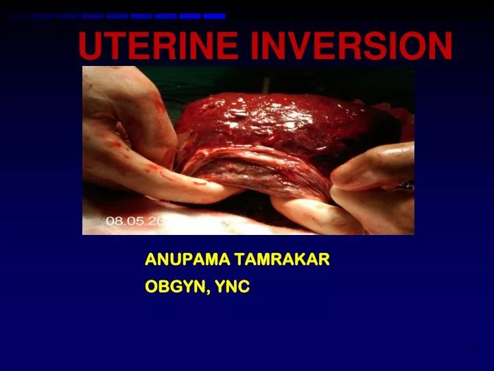
UTERINE INVERSION. ANUPAMA TAMRAKAR OBGYN, YNC. 1. Introduction. Prolapse of the fundus to or through cervix so that the uterus is turned inside out. Potentianlly life threatening complication of childbirth.

E N D
UTERINE INVERSION ANUPAMA TAMRAKAR OBGYN, YNC 1
Introduction Prolapse of the fundus to or through cervix so that the uterus is turned inside out.
Potentianlly life threatening complication of childbirth. • All most all cases occur after delivery/ C-Section in the third stage of labour.
Can occur even in the non-pregnant uterus in relation to the expulsion of an intrauterine tumour.
EPIDEMIOLOGY • Incidence varies widely. 1:2,000 to 1:23,000 deliveries.
TERMINOLOGY • Incomplete inversion describes an inverted fundus that lies within the endometrial cavity without extending beyond the external os. • Complete inversion describes an inverted fundus that extends beyond the external os.
A prolapsed inversion is one in which the inverted uterine fundus extends beyond the vaginal introitus. • A total inversion, usually non-puerperal and tumor related result in inversion of the uterus and vaginal wall as well.
CLASSIFICATION • 1st DEGREE The inverted fundus extend to but, not through the crevix.
2nd DEGREE The inverted fundus extend through crevix but remains in the vagina.
3rd DEGREE The inverted fundus coming out from vagina.
PATHOPHYSIOLOGY • Exact cause is unknown. • Principle behind its occurrence: Cervix must be dilated. Uterine fundus must be relaxed. • Many cases of acute uterine inversion results from mismanagement of third stage of labour in women who are already at risk.
If cervix is sufficiently dilated and the force of contraction sufficiently strong, the myometrial/ placental mass can be squeezed through it, resulting in complete inversion
In complete inversions once the fundus passes through the cervix, the cervical tissues function as a constricting band and edema rapidly forms. • The prolapsed mass then progressively enlarges and increasingly obstructs venous and finally arterial flow, contributing to the edema.
In chronic inversion serious tissue injury or necrosis is possible.
RISK FACTORS • Strong traction exerted on the umbilical cord due to adherent placenta. • Strong fundal pressure. • Fundal implantation of the placenta. 15
Endometritis /PID. D&C. Short umbilical cord. Certain drugs used as tocolysis. such as magnesium sulphate,nitroglycerine,turbutaline.
Precipitated labour • Prolong labour • Tumors-submucuos myomas . • Cervical incompetence. • Uterine anomalies( e.g unicornuate uterus). • Congenital or acquired weakness of the myometrium. 18
CLINICAL PRESENTATION Uterine inversion may present: ACUTE within 24 hours of delivery. SUB-ACUTE After 24 hours and up the 30th postpartum day. CHRONIC More than 30 days after delivery.
THE CLASSIC PRESENTATION: • Post-partum haemorrhage. • Sudden appearance of a vaginal mass. C Cardiovascular collapse ( varying degrees).
SYMPTOMS: • Pain in the lower abdomen. • Sensation of vaginal fullness with a desire to bear down after delivery of the placenta. • Vaginal bleeding unless the placenta is not separated.
SIGNS SHOCK • More commonly Neurogenic due to traction on the peritoneum and pressure on the tubes, ovaries, may be the intestine. • Parasympathetic effect of traction on the ligaments supporting the uterus and may be associated with bradycardia.
Shock: May be Hypovolaemic due to postpartum haemorrhage.
ABDOMINAL EXAMINATION • Cupping of the fundus in 1st & 2nddegree uterine inversion. • Absence of the uterus in 3rd & 4thdegree uterine inversion.
VAGINAL EXAMINATION • Soft, purple (dark bluish-red) mass in the vagina or vulva.
Diagnosing a first degree inversion is much more difficult. • Obesity can make diagnosis more difficult. • Chronic cases are unusual and difficult to diagnose. They may present with spotting, discharge and low back pain. Ultrasound may be required to confirm the diagnosis.
INVESTIGATIONS • Diagnosis is usually based on clinical symptoms and signs. • If not clinically very obvious, then USS & MRI is useful.
ULTRASOUND • Transverse image: A hypoechoic mass in the vagina with a central hypoechoic H-shaped cavity. • Longitudinal image: U-shaped depressed longitudinal groove from the unterine fundus to the centre of the inverted part. MRI
DIFFERENTIAL DIAGNOSIS OF UTERINE INVERSION • Uterine rupture. • Prolapse of uterine tumor (submucous fibroid). • Large endometrial polyp. • Passage of succenturiate lobe of placenta.
Inversion of uterus • Symptoms • Severe abdominal pain • Sudden cardiovascular collapse • Postpartum haemorrhage • Signs • Abdominal tenderness • Absence of uterine fundus on abdominal palpation • Lump in the vagina • Polypoidal red mass in the vagina with placenta attached
MANAGEMENT OF ACUTE & SUBACUTE UTERINE INVERSION AIMS Immediate treatment of Shock. Replacement /repositioning of the uterus.
TREATMENT OF SHOCK • Call for help. • IV line with two large bore IV cannulae. • Aggressive fluids replacement • Start resuscitation with normal saline or hartmann’s solution. 32
Blood transfusion. Analgesics. Use warm sterile towel to apply compression while preparing for the procedure. Insert a urinary catheter. 33
REPOSITIONING OF INVERTED UTERUS MANUAL REDUCTION. Sterile procedure. Form a fist or grad the uterus and push it through the cervix of a lax uterus towards the umbilicus to its normal position. Use the other hand to support the uterus. (Johnson maneuver)
Use of tocolytics to allow uterine relaxation. Nitroglycerin (0.25-0.5 mg) intravenously over 2 minutes. Terbutaline 0.1-0.25mg slowly intravenously. Magnesium sulphate 4-6 g intravenously over 20 minutes. Use of general anaesthesia: halothane. 35
O’SULLIVAN HYDROSTATIC METHOD. PRE-REQUISITES An assistant Long tube(2m) with a large nozzle Water reservoir/warm saline(2-5l).
PROCEDURE Trendelenburg position. Place the nozzle of the tube in the posterior fornix. An assistant start the douche with full pressure (at least 2m high) Fluid escape is prevented by blocking the introitus by using the labia and operator’s hand. The fluid distend the vagina, relieves the mild cervical constriction and result in correction or replacement of the inverted uterus. 37
NEW TECHNIQUE (ogueh and ayida) • Attaching the IV tubing to silicone cup used in vacuum extraction. By placing the cup in the vagina, an excellent seal is created.
AFTER REPOSITIONING • Discontinue uterine relaxant/general anaesthesia. • Start infusion of oxytocin or ergot alkaloids • Continue fluid and blood replacement • Bimanual uterine compression and massage are maintained until the uterus is well contracted and hemorrhage is ceased.
Remove placenta if retained following replacement of the inverted uterus. Careful manual exploration to rule out the possibility of genital tract trauma. Antibiotics . Adequate analgesics. Oxytocics\ergot are continued for at least 24 hrs. Monitor closely after replacement to avoid re-inversion
MANAGEMENT OF CHRONIC UTERINE INVERSION Surgical intervention. Abdominal route Vaginal route
Abdominal route • Huntington’s procedure • Haultain's procedure
HUNTINGTON PROCEDURE Locate the cup of the uterus formed by the inversion Dilate the constricting cervical ring digitally Stepwise traction on the funnel of the inverted uterus or the round ligament is given with Allis forceps Reapplied progressively as fundus emerges
(A) Obstetric ventouse applied on the inverted uterine fundus.(B) Reduction of the inverted uterus after traction with the ventouse.Instead of allies forceps alternativelt vacuum cup can be used in HUNTINGTON PROCEDURE
HAULTAIN’S PROCEDURE Incision is made in the posteriorly through the cervix, relieving cervical constriction to increase the size of the ring and allowing traction on the round ligament for the replacement of uterus with subsequent repair of incision from inside the abdomen 49
Vaginal route • Spinellis’s method • Kustner’s method • Hysterectomy 50