Download
1 / 19
220 likes | 913 Views
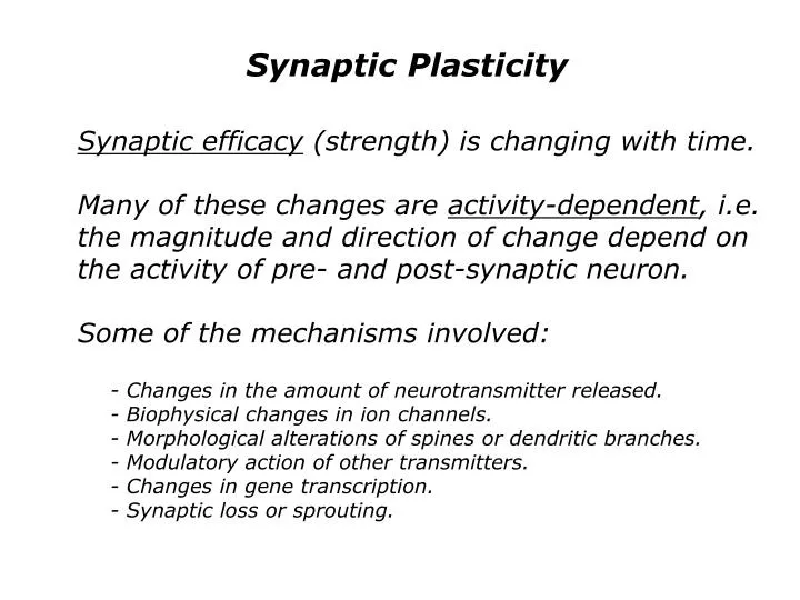
Synaptic Plasticity. Synaptic efficacy (strength) is changing with time. Many of these changes are activity-dependent , i.e. the magnitude and direction of change depend on the activity of pre- and post-synaptic neuron. Some of the mechanisms involved:.

E N D
Synaptic Plasticity Synaptic efficacy (strength) is changing with time. Many of these changes are activity-dependent, i.e. the magnitude and direction of change depend on the activity of pre- and post-synaptic neuron. Some of the mechanisms involved: - Changes in the amount of neurotransmitter released. - Biophysical changes in ion channels. - Morphological alterations of spines or dendritic branches. - Modulatory action of other transmitters. - Changes in gene transcription. - Synaptic loss or sprouting.
Hebb’s Postulate “When an axon of cell A is near enough to excite a cell B and repeatedly and persistently takes part in firing it, some growth process or metabolic change takes place in one or both cells such that A’s efficiency, as one of the cells firing B, is increased.” Donald Hebb, “Organization of Behavior”, 1949
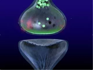
Animal Models of Plasticity Long-Term Potentiation (LTP) Cross-section of the hippocampus: Cajal’s drawing
Animal Models of Plasticity Brain slice preparation of the hippocampus:
LTP Typical LTP experiment: record from cell in hippocampus area CA1 (receives Schaffer collaterals from area CA3). In addition, stimulate two sets of input fibers.
LTP Typical LTP experiment: record EPSP’s in CA1 cells (magnitude) Step 1: weakly stimulate input 1 to establish baseline Step 2: give strong stimulus (tetanus) in same fibers (arrow) Step 3: continue weak stimulation to record increased responses Step 4: throughout, check for responses in control fibers (input 2)
LTP LTP is input specific. LTP is long-lasting (hours, days, weeks). LTP results when synaptic stimulation coincides with postsynaptic depolarization (achieved by cooperativity of many coactive synapses during tetanus). The timing of the postsynaptic response relative to the synaptic inputs is critical. LTP has Hebbian characteristics (“what fires together wires together”, or, in this case, connects together more strongly). LTP may produce synaptic “sprouting”.
The NMDA Receptor • At the resting potential (postsynaptic neuron), glutamate binds to the NMDA channel, the channel opens, but is “plugged” by a magnesium ion (Mg2+). • Depolarization of the postsynaptic membrane relieves the magnesium block and the channel open to allow passage of sodium, potassium and calcium.
The Associative Nature of LTP Old(er) view: Associative requirement is mediated by the voltage-dependent characteristics of the NMDA receptor. New discovery (1994): Active conductances in dendrites mediate back-propagation of AP’s into the dendritic tree.
Spike-Timing Dependent Plasticity Basic Idea: Change in synaptic strength depends on the precise temporal difference between pre- and post-synaptic neuronal firing (causality!).
The Neuron: Integrator or Coincidence Detector? Synchronous inputs really matter!
Data Analysis in Neurophysiology Spike train data sets: Neuron in MT Colby and Duhamel, 1991
Data Analysis in Neurophysiology Neuron in IT (object selective) Desimone et al., 1984
Data Analysis in Neurophysiology Neurons in V1 (orientation selective) PSTH (firing rate) Auto-Correlation Shift Predictor Cross-Correlation Engel et al., 1991
Neural Coding Rate coding versus temporal coding One major mechanism of how neurons encode information is through their firing rate (number of AP’s per second). – Example: orientation selectivity. Another major mechanism is synchronization (AP’s occurring together in time). – Example: perceptual grouping. Synchrony could affect other neurons (e.g. through spatial summation – see unit 1).
Computational Neuroscience Components of (most) neural models: - Units and connections - Inputs and outputs - Activation function - Learning rule
“Why the Mind is in the Head” “Why is the mind in the head? Because there, and only there, are hosts of possible connections to be formed as time and circumstance demand. Each new connection, serves to set the stage for others yet to come and better fitted to adapt us to the world, for through the cortex pass the greatest inverse feedbacks whose function is the purposive life of the human intellect.” Warren S. McCullogh, Hixon Symposium 1951.