Download
1 / 56
600 likes | 1.11k Views
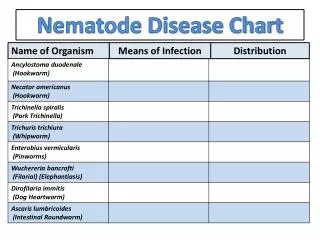
The intestinal nematodes Ascaris Hook worms Pin worm Whip worm. The blood- and tissue dwelling nematodes The filaria Trichinella . Nematode. Ascaris 蛔虫. Ascaris lumbricoides 似蚓蛔线虫. Introduction. The first representative and the most common intestinal parasite

E N D
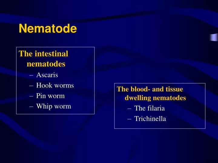
The intestinal nematodes Ascaris Hook worms Pin worm Whip worm The blood- and tissue dwelling nematodes The filaria Trichinella Nematode
Ascaris蛔虫 Ascaris lumbricoides 似蚓蛔线虫
Introduction • The first representative and the most common intestinal parasite • Cosmopolitan in distribution • Rural > urban • Children > adults
Morphology Adult Looks like an earthworm Female (20-35 cm); Male (12-30 cm) 3 lips which carry minute teeth
A pair of female and male worms of A. lumbricoides. Notice the vulvar waist(arrow)of the female worm and the coiled end of the male worm.
A scanning electron micrograph of Ascaris showing the three prominent “lips”
Egg • Fertilized egg • Unfertilized egg
Egg shell Ovum Albuminous layer A. lumbricoides, fertilized egg (6050 micrometer)
Living site Adults in small intestine HOST MAN Migration Larvae migrate though the lungs Diagnostic stage Undeveloped eggs in feces Method of infection Infective eggs are ingested Infective stage Eggs embryonate in soil by 2-3 wks
Symptomatology Larva Pneumonitis Asthma attacks Loeffler’s syndrome
Adult • The presence of few worms may be asymptomatic (85%) • The most common symptoms are vague abdominal pain • Large numbers of worms may cause malnutrition and present signs and symptoms of obstruction
Migration of adult worms may cause signs and symptoms of perforation, peritonitis, appendicitis or extrahepatic biliary obstruction.
Cross section of a liver specimen contains many adult worms of A.lumbricoides obstructing the intrahepatic and extrahepatic bile ducts.
A large mass of Ascaris lumbricoides that was passed from the intestinal tract. The ruler at the bottom of the image is 4 cm (about 1.5 inches) in length.
An autopsy specimen shows intestinal obstruction by many adult worms of A.lumbricoides. Notice the markedly distended intestinal loop, the thin intestinal wall with hemorrhage and worms protruding from the perforated wound.
Peritonitis caused by intestinal perforation due to Ascaris Resected bowel and the adult female from the peritoneal cavity
Diagnosis • Microscopic identification eggs in the stoola direct wet mount examination of the specimen (200,000 eggs/female/day) • Macroscopic identification of adults passed in stool or through the mouth or nose
Epidemiology • Worldwide distribution, throughout the temperate and tropical areas • 1,000,000,000 people in the world • 40% population in Africa and Asia • 600,000,000 in China (1992)
Treatment • Albendazolea single oral dose of 400 mg • Mebenazole100 mg orally twice daily for 3 days
Prevention • Avoid contacting soil that may be contaminated with human feces • Do not defecate outdoors • Dispose of diapers properly
Wash hands with soap and water before handling food • When traveling to areas where sanitation and hygiene are poor, avoid water or food that may be contaminated • Wash, peel or cook all raw vegetables and fruits before eating
Ancylostoma duodenale十二指肠钩口线虫Necator americanus美洲板口线虫
Morphology • Adult • Cylindrical with the head bent sharply backwards • Males are smaller than the females and possess a bursa at their posterior end
Scanning electron micrograph of the oral opening of Ancylostoma duodenale, another species of human hookworm. Note the presence of four cutting "teeth," two on each side.
Adult mouthpart of Necator americanus Note : The large buccal capsule is open dorsally with one pair of cutting plateteeth.
Bursa of hookworms Lift A. duodenale; Right N. americanus
Enterobius vermicularis adult female 0.8-1.3cm.in length , spindle-shaped with a long thin sharply tail. The greater part of the body is occupied by the uterus filled with eggs.
Egg (indistinguishable between the 2 species) • Median size (like the ascaris egg) • Elliptical • Transparent • Thin shell • 4-cell stage when discharge
Enterobius vermicularis egg. Note the thick shell and characteristic shape; approximate length = 55 µm.
Egg < Ascaris egg Non-symmetrical ellipse; D shaped Transparent Thick transparent shell Tadpole-like embryo when discharged
Life Cycle • Host -man • No intermediate host 钩虫: Egg Larva (free-living) Larvae migrate fromskin to the lungs 蛲虫:Egg takes 6 hours to be infective stage 鞭虫: Similar to Ascaris but no pulmonary migration . There are reservoir hosts.
Parasitic site: 蛔虫: small intestinal 钩虫: upper small intestine; Duodenum, jejunum 蛲虫: colon Gravid female adult deposits its eggs on the anus and perianal skin. 鞭虫: ileo-caecal region
Infection stage 蛔虫: infective egg 钩虫: infective larva 蛲虫: infective egg ( Egg takes 6 hours to be infective stage) infective mode: Anus-Hand-Mouth Auto-infection/Cross-infection 鞭虫: infective egg
Pathogenic stage: 蛔虫: adult worm /larva 钩虫:adult worm Digestive disturbances /Allotriophagy Microcytic hypochromic anemia (sucking, oozing, discharging)A.d. 0.15-0.4ml/d; N.a. 0.02-0.1ml/d larva:Dermatitis ‘ground itch’ Pneumonitis and Bronchitis