Download
1 / 40
400 likes | 966 Views
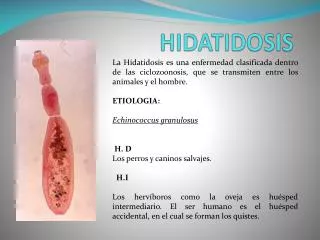
HIDATIDOSIS. Dr. César Náquira. HIDATIDOSIS Definición. Zoonosis parasitaria causada por la larva ( hidatide ) de Echinococcus granulosus , cestodo del intestino del perro ( hospedero definitivo )

E N D
HIDATIDOSIS Dr. César Náquira
HIDATIDOSISDefinición Zoonosis parasitaria causada por la larva (hidatide) de Echinococcus granulosus, cestodo del intestino del perro ( hospedero definitivo) El ganado y animales hervíboros son los hospederos intermediarios habituales (contienen la larva). El ser humano es un hospedero intermediario accidental Ampliamente distribuida, principalmente en zonas ganaderas
Echinococcus granulosus: adultoen vellosidades intestinales del perro
HIDATIDOSISEchinococcus granulosus: adultoen vellosidades intestinales del perro escolex Proglotida grávida
HIDATIDOSIS EN EL PERÚINFECCIÓN CANINA% INFECCIÓN SIERRA CENTRAL (Junín) 8 - 46 SIERRA SUR (Puno) 31.3 - 37 LIMA (capital-zona urbana) 2 - 3.4 AREQUIPA (Sur-zona urbana) 8 - 48.2
Quistes hidatídicos en pulmón ganado humano
Quiste hidatídico: corte histológico Vesícula prolígera Membrana prolígera Cutícula Adventicia Hígado normal
HIDATIDOSIS EN EL PERÚINFECCIÓN ANIMAL% INFECCIÓN SIERRA CENTRAL SIERRA SUR VACUNOS 50 16 - 69 OVINOS 26.7 13.2 - 46.96 CAPRINOS 2.7 5.1 PORCINOS 2.8 9.1 AUQUÉNIDOS ..... 2.1 - 8.32
HIDATIDOSIS EN EL PERÚINFECCIÓN HUMANA Tasa x 100.000 Necropsias (Morgue central de Lima)190 Necropsias (Hospital de Lima) 530 CASOS NOTIFICADOS (MINSA) 605 (1983) 2000 (2002) ZONA ENDÉMICA Abreugrafía – Junín 3190 (1973) Serología (arco 5)- Junín 5800 (1994) Serología (inmunoblot)- Junín 15000 (2001)
HIDATIDOSISDIAGNÓSTICO DE LABORATORIO EXAMEN DIRECTO: OBSERVACIÓN DE ESCOLEX, GANCHOS EN ESPUTO, VÓMICA O PUNCIÓN EXAMEN INDIRECTO: BÚSQUEDA DE ANTICUERPOS EN SUERO SANGUINEO Pruebas de tamiz: HAI, ELISA, partículas de latex Poseen alta sensibilidad y baja especificidad Pruebas de elección: IEF, DD5 y Western Blot BÚSQUEDA DE ANTÍGENO EN LÍQUIDO DE PUNCIÓN
INMUNOELECTROFORESIS Anticuerpo Antígeno Arco 5
Doble Difusión del arco 5 (DD5) Antígeno Suero problema Suero problema + + Anticuerpo - + - -
INMUNOBLOT I. SDS-PAGE
INMUNOBLOT Banda 21-31 kd
CISTICERCOSIS Dr. César Náquira
CISTICERCOSISDEFINICIÓN Zoonosis parasitaria causada por la larva (cisticerco) de Taenia solium. Hospedero definitvo de Taenia solium: hombre tiene el parásito adulto Hospedero intermediario habitual: cerdo tiene la larva (cisticerco) Hospedero intermediario accidental: el hombre tiene la larva (cisticerco)
CISTICERCOSIS: diagnóstico Antecedentes epidemiológicos Imágenes: Rayos X, Ecografía, TAC, resonancia magnética. Laboratorio: Sangre: búsqueda de anticuerpos: ELISA, WESTERN BLOT Heces: búsqueda de huevos de Taenia solium