Download
1 / 54
590 likes | 900 Views
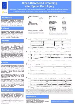
Using Holter ECG and Heart Rate Variability to Detect Sleep-Disordered Breathing. Phyllis K Stein, Ph.D. Heart Rate Variability Laboratory Washington University School of Medicine St. Louis, MO. Background.

E N D
Using Holter ECG and Heart Rate Variability to Detect Sleep-Disordered Breathing Phyllis K Stein, Ph.D. Heart Rate Variability Laboratory Washington University School of Medicine St. Louis, MO
Background When patients with sleep-disordered breathing have an event, there is an autonomic arousal associated with a brief awakening, they then resume normal breathing, and fall back asleep. This repeated awakening is associated with a repeated increase in heart rate which return to baseline when the patient falls back asleep.
Heart-Rate-Based Graphical Method for Detecting Sleep-Disordered Breathing 1. Sequence of unedited beat-to-beat R-R (or preferable edited N-N) intervals. 2. Convert R-R intervals to instantaneous HR (60,000/R-R interval in ms). 3. Plot tachogram of HR vs. time on 6 parallel 10-min plots (one hr/page).
0-100 bpm “x-axis” Tachogram Axes • x-axis = time in minutes (0-10 minutes) • y-axis for each 10-min plot is H (0-100 bpm in 5 cm) • “x-axis” is mean HR for that 10-min segment
To bed Sleep Onset in a Patient Without OSAHS
Onset of OSAHS Patient falls asleep
Tachograms From the Computers In Cardiology Sleep Apnea Contest • Data based on R-R intervals using simple QRS detection algorithm andnot edited. • 35 tachograms blindly scored for OSA, no OSA and indeterminate. # each category known. • Graphical method, 1 pair wrong, severe sleep-disordered breathing but hypopneas not OSA.
Brady-tachy pattern not seen CVHR Subject 2
CVHR Subject 5 Tachycardia during OSA
Probable change in position resulting in OSA CVHR and Normal Sleep or Quiet Rest Subject 9
Change in position terminates apnea Apnea Appears to be Positional in Subject 23
Probable change in position-apnea more severe earlier CVHR Subject 30
Magnitude of RSA declines during some but not all events Severe Sleep Apnea Subject 31
Probable change in position or sleep stage. RSA is reduced. Severe Sleep Apnea Subject 32
Tachogram Evaluation • Identify epochs of CVHR (cyclic variation of heart rate) • Quantify CVHR by by total number of minutes (to nearest 30s) with CVHR. • If CVHR is predominant, no need to quantify.
CVHR Definition • At least 3 consecutive cycles of rising and falling heart rate. • A visible rise in heart rate (5 bpm). • A return to baseline. • Each cycle 10 s duration. • At least 20s but less than 2 min between cycles.
CVHR Criteria for Significantly Abnormal Sleep • 20% of time in CVHR of any type • High amplitude regular CVHR pathomnemonic for OSA • Lower amplitude or irregular CVHR may be associated with apneas, hypopneas, periodic limb movements or arousals for no apparent reason.
Results of Sleep Lab Validation of CVHR Tachogram Method • 100% detection of significantly abnormal sleep. • High amplitude regular CVHR always sleep apnea. • Lower amplitude or irregular CVHR could be apneas or hypopneas or leg movements, a mixture or arousals for no apparent reason. • Non-diagnostic for flat tachograms (extremely low HRV) or atrial fibrillation.
Heart Rate Patterns on Tachograms Can Detect More Than Just Sleep Apnea
O2 Sat = 65% Irregular Low Amplitude CVHR HR Patterns During Severe De-Saturation
Blown Up Section of Prior Tachogram Showing RSA During Cheyne-Stokes Respiration
Power Spectral Analysis of Heart Rate Variability to Detect Sleep-Disordered Breathing
HRV power spectral plot quantifies the underlying periodicities in heart rate. • CVHR is a periodic change in heart rate which should be reflected in the HRV power spectrum
HF Peak Due to RSA Normal-Appearing Nighttime Power Spectral Plot
Patient falls asleep Onset of OSAHS
VLF Peak Associated with Sleep Apnea HF Peak Due to RSA 0 0.8 Hz Power Spectral Plot for Previous Tachogram Showing OSAHS Pattern
0 0.8 Hz Power Spectral Plot for Previous Tachogram Showing HRV Pattern for Central Apneas VLF Peak Associated with Central Apneas Little or no HF power
O2 Sat =65% Irregular Low Amplitude CVHR HR Patterns During Severe De-Saturation
VLF Peak Associated with OSAHS Diffuse HF Peak Reflecting Irregular Respiration or Heart Rate Pattern 0 0.8 Hz Power Spectral Plot for Previous Tachogram
2-Min Averaged HRV Pattern for Cheyne-Stokes Respiration Hard to see CSR peak