Download
1 / 26
260 likes | 478 Views
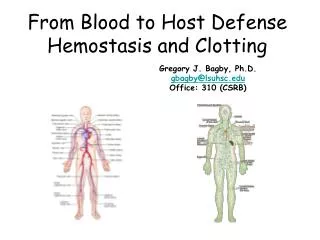
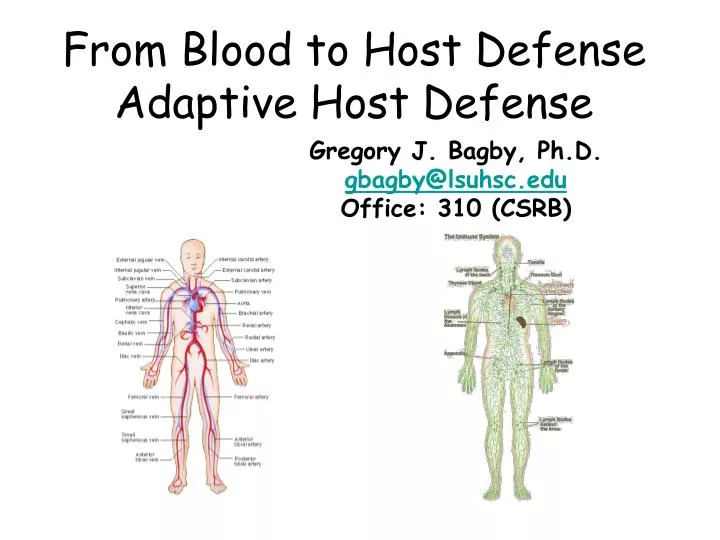
From Blood to Host Defense Adaptive Host Defense. Gregory J. Bagby, Ph.D. gbagby@lsuhsc.edu Office: 310 (CSRB). From Blood to Host Defense. Blood Components and function Hemostasis and clotting The host defense system General overview Innate immune system pathogen recognition

E N D
From Blood to Host Defense Adaptive Host Defense Gregory J. Bagby, Ph.D. gbagby@lsuhsc.edu Office: 310 (CSRB)
From Blood to Host Defense • Blood • Components and function • Hemostasis and clotting • The host defense system • General overview • Innate immune system • pathogen recognition • inflammatory response • Adaptive immune system • Humoral immune system and antibodies • Cell-mediated immune system
Adaptive Host Defense System • Overview of the adaptive immune system • Functions of B and T lymphocytes (cells) • B lymphocyte receptors and antibodies • Ag-binding sites diversity • T lymphocyte receptors • Antigen presentation to B and T cells • B cells – Ag can bind to B cell receptor • T cells – Ag must be presented to the T cell receptor • Development of immune tolerance • Antibody-mediated immune response • Defenses against virus-infected cells and cancer cells • Role of NK cells and macrophages
Overview of Adaptive Host Defense System • Lymphocytes are the cells of the adaptive immune system • Any molecule that triggers an adaptive response against itself or a cell bearing it, is called an antigen (immunogen) • Protein, protein fragment, polysaccharide • Host recognizes as non-self • Highly specific and adaptive • Lymphocyte organs – Where do the lymphocytes hang out? • Lymphocyte origins • Three stages of acquired response • Recognition – one lymphocyte, one antigen • Activation – lymphocyte clonal expansion • Attack – eliminate antigen or kill antigen-bearing cell.
Anatomy of the Adaptive Immune System: Lymphoid Organs • Primary lymphoid organs: • Supply secondary lymphoid organs with mature lymphocytes • Bone marrow • Thymus • Secondary lymphoid organs: • Areas where lymphocytes from 1° lymphoid organs divide and reside • Lymph nodes, tonsils • Spleen • Mucosal-associated lymphoid tissue (MALT) – intestines, respiratory, genital & urinary
Anatomy of the Adaptive Immune System – Lymphatics and Lymph Nodes • Function • Filter particulates and microbes • Antigen presentation • Components • Cortex – B cell rich • Paracortex – T cell rich • Accessory cells (macrophages, dendritic cells, others) located in each • Medulla - Macrophages
Activated by antigen Maturation & Differentiation of Lymphocytes Pluropotentstem cell Bone marrow Lymphoid Myeloid Mature B cell Naïve T cell Secondary lymphoid organ Plasma cell Thymus Antibodies (maturation) NaïveHelper T cell Effector&MemoryCells NaïveCTL cell
Recognition, Activation and Attack Recognition Antigen Free Presented B cell Helper T cell Cytotoxic T cell Activation Cytokines Cytokines Plasma cell Antibodies Attack Guide phagocytes, complement, and NK cells to free antigen and Ag-bearing cells Attack antigen-bearing cells
Lymphocytes Are the Cells of the Adaptive Immune System • B lymphocytes (cells): • Naïve B cells – B cell receptor • Memory B Cells – B cell receptor • Plasma Cells – Ab secreting • T lymphocytes (cells): • T helper cells • Cytotoxic T cells • Naïve T cells (helper and CTL) • Effector T cells (helper and CTL) • Memory T cells (helper and CTL) • T regulatory cell
Functions of B Cells • B cells participate in antibody-mediated responses (humoral) • Extremely wide diversity of molecular targets. • B cells recognize antigens via B cell receptor. Each B cell has a unique receptor for a specific antigen (Ag) • Major defense against bacteria, viruses in extracellular fluid • B cells differentiate into plasma cells whichsecrete antibodies (Ab) • Secreted Ab enter the blood. • If form an Ab-Ag complex lead to neutralization and/or removal of the Ag
B Cell Receptors Are Immunoglogulins (Ig) Specific antigenbinding sites • B cell receptor - copies of specific Igon its plasma membrane • Glycoprotein acts as receptor for its antigen • Receptor also called an Ig • Not secreted therefore B cell receptor not an antibody • B cell receptor and antibodies are Ig and composed of 4 interlinked polypeptide chains. • 5 major Ab classes – IgA, IgD, IgE, IgG, IgM • Ag-binding site - variable region (millions of unique amino acid sequences) each capable of binding one specific Ag • Fc stem – identical within Ab classes • Plasma cells are clones of B cells with identical variable regions Light chain Variable end Heavy chain Constant end Fcstem
B Cell Receptor Diversity • Human genome contains about 200 genes that code Ig • How does body produce millions of different Ag-binding sites? • Answer – type of genetic recombination unique in developing lymphocytes • Process requires enzyme only found in developing lymphocytes to perform the task during development • Variable region cut into segments and randomly rearranged • Varies from B cell to B cell resulting in millions of different unique sequences each capable of binding to a single Ag
Functions of T Cells • T cells play a variety of roles to include cell-mediated as opposed to humoral responses. • Different kinds of T cells (CD3+) • Helper T cells – CD4+ • Cytotoxic T cells – CD8+ • Regulatory T cells – CD4+ • Cytotoxic T cells are attack cells • Travel in blood and tissues to seek out and bind to antigen-bearing target cells • Kill target cells by secreting chemicals • Cancerous or infected cells are killed by CD8+ cells • Helper T cells assist in activation and function of B cells, CD8+ cells and macrophages (dendritic cells) – Th1, Th2, Th17 • Regulatory T cells believed to suppress activities of B and CD8+ cells
Antigen Recognition by T Lymphocytes Requires Presentation to the T cell Receptors • T cell receptor: Two-chained proteins are similar to B cell Ig on cell surface • Each T cell has receptor specific for one particular Ag. Similar variable region to B cell receptor and Ab • As in B cells, multiple DNA rearrangement result it millions of distinct T cell clones • T cell receptor remains embedded in plasma membrane of all T cells • T cell receptor can only bind to its Ag that is “presented” to it in combination with a bodies own plasma membrane proteins (self proteins) • Group of proteins collective called major histocompatibility complex (MHC) or human leukocyte associated antigens (HLA Ag) • No two humans, except identical twins, have the same MHC genes • Markers of biological self
Major Histocompatibility Complex (MHC) • MHC proteins are called “restriction elements” because ability of T cell receptors to identify Ag is restricted to the Ag complexed to an MHC protein • Two classes of MHC: • Class I: • On surface of virtually all nucleated cells • Required for Ag presentation to cytotoxic T cells • Class II: • Only on surface of macrophages, macrophage-like cells, B cells, and dendritic cells. • Required for presentation to helper T cells. • Ag are recognized by T cells only when complexed with MHC of an antigen presenting cell (APC)
Additional Requirements for APC to Present Ag to Helper T Cells • Ag complexed with Class II MHC • Costimulus with nonantigenic matching proteins • APC secretion of IL-1 and TNF • Activated helper T cell then secretes cytokines with autocrine and paracrine effects on nearby cells • B cells • Cytotoxic T cells • NK cells • Macrophages
Development of Immune Tolerance • Diverse lymphocyte receptors result from random DNA cutting and recombination. Recognize both self and nonself molecules. • Immune tolerance occurs in early life resulting in lymphocytes that lack immune responsiveness to self molecules/proteins. • Clonal deletion via programmed cell death (apoptosis) • Clonal inactivation or anergy– render cells nonresponsive • Self molecule presentation occurs in thymus (T cells) and bone marrow (B cells) during cell development • Ag presentation to helper T cells occurs without costimulation which results in cell death or permanent inactivation. • Immature B cells only express IgM. If they bind self molecule during in bone marrow they undergo clonal deletion
Sequence of Events in Antibody-Mediated Immunity Against Bacteria • Bacterial Ag bind to B cell receptor on B cell in secondary lymphoid organ. • Simultaneously and in the same microenvironment, B cells, and possibly APCs, present Ag complexed with MHC II to helper T cells via T cell receptor. • Helper T cell secrete IL-2, IL-4, etc. • Stimulates helper T cell to proliferate • Stimulates Ag-bound B cells to proliferate and differentiate into B memory cells, plasma cells. • Plasma cells secrete antibodies specific for Ag that initiated the process. • Antibodies circulate and combine with Ag on the surface of bacteria or free in the extracellular fluid or possibly on cells. • Ab-Ag complex causes conformational change in the Fc-stem • Facilitates phagocytosis by neutrophils and macrophages • Activates the complement system which also facilitates phagocytosis and directly kills bacteria via MAC • Induces antibody-dependent cellular toxicity via NK cells.
Antibody Production Kicks into High Gear with 2nd Exposure to Ag Memory cells, produced along with plasma cells during the first infection, quickly generate large numbers of antibody molecules during a second infection.
Consequences of Antibody Binding to Antigen • Antibodies do not kill on their own • Can “neutralize” cell-free viruses, protein or toxins • Ab link microbes to actual killing mechanisms • Opsonization for phagocytosis • Activation of the complement system – MAC attack • Antibody-dependent cellular cytotoxicity • NK cells bind to Ab-antigen complex
Sequence of Events in Cytotoxic T Cell-Mediated Immunity Against Cells Infected with Virus • Viral Ag complexed to MHC I is presented (binds) to T cell receptor on CTL • Simultaneously, viral processed by APC and its Ag:MHC II complex presented to helper T cell. • Helper T cell secretes cytokine like IL-2 & IFNγ which stimulate viral Ag activated cytotoxic T cells • CTL proliferate/differentiate into effector and memory CTL. • Helper T cells proliferate/differentiate into effector and memory helper T cells • CTL attack and kill virus infected cells via secreted proteins (perforin). • Liberated virus opsonized (Ab or complement) and phagocytized (macrophages or neutrophils)
Cell-Mediated Host Defense against Virus Infected Host Cells Virus infected cells present a viral antigen to be attacked by cytotoxic T-cells, which release a protein (perforin) that results in the perforation of the infected cell, leading to the leakage of its contents and cell death.
Role of NK Cells and Macrophages in the CTL Attack • Activated NK cells and macrophages participate in destroying virus-infected or cancer cells tagged with CTL • IL-12 and IFN-γ are the signals for activation • System is a positive feedback system because activated NK cells secrete IFN-γ • Both Secrete toxic chemicals • Macrophages - phagocytosis