Download
1 / 66
1.38k likes | 2.9k Views
Short notice on non-vascular INTERVENTIONAL RADIOLOGY. Presented by Hisham S. Wahba Ass. Lecturer of radiodiagnosis National Cancer Institute, Cairo University. INTRODUCTION.

E N D
Short notice on non-vascular INTERVENTIONAL RADIOLOGY Presented by Hisham S. Wahba Ass. Lecturer of radiodiagnosis National Cancer Institute, Cairo University
INTRODUCTION • Interventional radiologists are doctors who specialize in minimally invasive, targeted treatments that have less risk, less pain and less recovery time compared to open surgery. • They use their experience to map out the procedure tailored to the individual patient to treat diseases at the site of the illness nonsurgically. • Interventional radiology is a recognized medical specialty by the America Board of Medical Specialties.
INTRODUCTION • GOAL -------> simplify treatment in a way that minimizes patient discomfort, renders general anesthesia unnecessary, lowers the incidence of morbidity and mortality, and decreases the length and cost of hospitalization. • Special procedures can replace surgery (embolization of bleeding ulcers). • Others can complement surgery (postoperative abscess drainage). • Certain procedures can be used in the management of conditions for which there is no surgical solution (selective chemotherapy).
INTRODUCTION DIAGNOSTIC
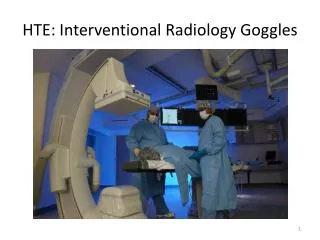
Interventional radiology operating theater. NON-VASCULAR TECHNIQUES
PERCUTANEOUS BIOPSY Fine Needle Aspiration Cytology (FNAC) True Cut Core Biopsy
FNAC and True Cut Core BiopsyIndications • Presence or absence of disease. • Nature of disease (neoplastic, inflammatory, infectious). • Extent of disease.
FNAC and True Cut Core BiopsyContraindications • Abnormal coagulation profile ---Elevated prothrombin time (PT). ---Elevated partial thromboplastin time (PTT). ---Depressed platelet counts. ---Low prothrombin concentration (PC).
FNAC and True Cut Core BiopsyPatient preparation • Informed consent. • Normal bleeding profile. • Start clear liquid diet, the night before the procedure. • Anxiolytic agents (anxious patients, severely painful biopsies like bone, infants). • Sterilize the puncture site, drape the surrounding area.
FNAC and True Cut Core BiopsyEquipment • FNAC ----- spinal needles (20 – 22 Gauge) • True Cut Core Biopsy ----- true cut biopsy needle (unicut 16G 15cm), gun biopsy needle (speedybell 18G 20cm). 3 1 2
FNAC and True Cut Core BiopsyTechnique • Ultrasound or CT guided. • Precise lesion site. • Apply local anaesthetic (1 % lidocaine) subcutaneous (True Cut Biopsy). • Insert needle under guidance. • Cytopathologist at biopsy procedure can minimize number of passes.
FNAC and True Cut Core BiopsyComplications • Pain and discomfort. • Hemorrhage (heamatoma). • Pneumothorax (lung biopsy).
Percutaneous Abscess DrainagePatient preparation • Normal bleeding profile. • Fasting for at least 6 hours. • Sterilize the puncture site, drape the surrounding area.
Percutaneous Abscess DrainageTechnique • Subcutaneous lidocaine 1 % injection. • Image guided needle placement. • Guide wire introduced through sheathed needle. • Catheter (pigtail) advanced over guide wire. • Catheter fixed in place.
Percutaneous NephrostomyIndications • DECOMPRESSION ---Stone ---Stricture ---Tumor • URINOMA
Percutaneous NephrostomyEquipments • Needle (PTC catheter set) 19 G 25 cm • Guide wire 0.38’’, 150 cm (Teflon). • Dilators, 6F, 8F, 10F and 12F. • Pigtail catheter (nephropur) 8F.
Percutaneous NephrostomyPatient preparation • Labs: CBC, Coagulation Profile, serum Creatinine, BUN, urine culture and ECG. • Ultrasound: determine location of kidney, degree of hydronephrosis. • Patient sedation if anxious. • Skin sterilization.
Patient lies in prone position. 1 % lidocaine injection. Needle inserted under US guidance. Tocar withdrawn. Contrast media injected through needle sheath (opacify pelvi-calyceal system) under fluoroscopy. Guide wire introduced through needle sheath. Sheath withdrawn. Dilator inserted successively (6F12F). Catheter advanced upon guide. Catheter fixed insitu. Percutaneous NephrostomyTechnique
Percutaneous NephrostomyCatheter Maintenance • Catheter skin site clean and dry. • Exchange and maintain bag in aseptic manner. • Bag always lower than kidney to ensure proper gravity drainage. • Call radiologist if any symptoms or signs of drain obstruction occurs. • Drains changed at 2 to 3 months intervals.
Percutaneous Nephrostomycomplications • Occlusion of nephrostomy drain. • Displaced nephrostomy catheter. • Hemorrhage. • Sepsis. • Urinoma.
PERCUTAEOUS BILIARY DRAINAGEEQUIPMENT • Needle (PTC catheter set) 19 G 25 cm • Guide wire 0.38’’, 150 cm (Teflon). • Dilators, 6F, 8F, 10F and 12F. • Pigtail catheter 8F.
Cancer pancreas. Gallstones. Strictures Cholangiocarcinoma. Sclerosing cholangitis. Cancer gall bladder, CBD, ampulla. Liver abscess. Duodenal diverticulum. Caroli’s disease. Retroperitoneal fibrosis. Parasites. PERCUTAEOUS BILIARY DRAINAGE Causes of bile duct obstruction
PERCUTAEOUS BILIARY DRAINAGEPatient preparation • Normal coagulation factors (PC > 75%). • Clear fluids after midnight. • Fasting at least 6 hours. • Prophylactic antibiotic for 12 to 24 hrs. • Written consent.
Skin sterilization. Local anesthetic (1% lidocaine). 3 mm stab to skin made by surgical blade. Needle advanced parallel to table top from a point at Rt 9th intercostal space at midaxillary line. Small amount of contrast injected through needle while it is slowly withdrawn under fluroscopy. We can use US guidance as alternative to point to dilated biliary radicle. When contrast fills biliary tree, a guide wire is advanced through needle under screen till reaching the CBD. Dilators are applied successively (6F- 9F). Finally, pigtail catheter (external drain) is inserted. Catheter then fixed to skin PERCUTAEOUS BILIARY DRAINAGETechnique
PERCUTAEOUS BILIARY DRAINAGEComplications • Sepsis and bleeding. • Hemobilia. • Punturing extra-hepatic structures.
PERCUTAEOUS BILIARY DRAINAGEPrevention of Complications • Use 22 or 21 Gauge needles. • Take care not to puncture gall bladder and colon. • Keep contrast volume to a minimum to prevent rise in bile pressure. • Use potent antibiotic coverage before and during PBD. • Keep patient well-hydrated.
RADIOFREQUENCY ABLATION Liver Lung
Radiofrequency Ablation LiverPhysical background • Basics of RFA: • RF energy is an alternating current with a frequency of 10-900 MHz. RF waves have long WL and as such are of a very low energy.
Radiofrequency Ablation LiverPhysical background • RF energy effect on body tissues: • When RF electric field is applied to the body, the interaction losses in moving ions and water molecules at a frequency of the electric field, creating conduction current. • The friction and ionic agitation generate heat that is produced within the tissues near the electrode called “resistive heat”.
Radiofrequency Ablation LiverPhysical background • Induction of Coagulative necrosis: • The aim of tumour ablation therapy is to destroy the entire tumour by using heat to kill malignant cells and including 0.5-1 cm safety margin. • The aim for RF ablation is achieving and maintaining a 50-100ºC-temperature range throughout the entire target volume.
Radiofrequency Ablation LiverIndications • Disease confined to the liver, without evidence of vascular invasions or distal metastases. • Tumour size should be ideally smaller than 4 cm. • PC > 50% and a platelet count > 50,000/mm3. • No other site of metastases in colorectal carcinoma metastases.
Radiofrequency Ablation LiverApproaches • Percutaneous approach, is the least invasive, with minimal morbidity, can be performed on an outpatient, requires only sedation, and can be repeated. • Laparoscopic approach, • Open surgical approach, has associated morbidity and mortality of an open procedure and general anaesthesia, and the technique is a one shot therapy.
Radiofrequency Ablation LiverImaging interpretation & follow-up • AFP for HCC and CA19.9 for CR metastases are of limited value for assessing tumour response because patients with small tumors may have normal pretreatment levels. • So, the evaluation of therapeutic effect is based mainly on findings of imaging studies, which accurately reflect the efficacy of ablation. • The US shows hyperechoic area replacing the original lesion. This feature, however is unreliable for assessing the outcome of treatment.
Radiofrequency Ablation LiverImaging interpretation & follow-up • Spiral CT imaging: • Spiral CT is the standard imaging modalities for evaluating tumour response after RFA. • Successfully ablated tumours appear as hypodense areas on CT and do not enhance on the arterial and venous phases after contrast injection. • Spiral CT may show the presence of a peripheral enhancing halo surrounding the treated lesion.
Radiofrequency Ablation LiverImaging interpretation & follow-up • This halo is due to the hyperemia and the inflammatory reaction along the periphery of the ablated area. • This enhancing halo is depicted several days after treatment and usually disappears 1 month later. • A standard protocol for the follow-up of treated cases should include at least a spiral CT study of the liver every 3-4 months .
Radiofrequency Ablation LiverComplications • Bleeding. • Infection. • Biliary tract damage. • Liver failure. • Pulmonary complications. • Dispersive pad skin burns. • Hepatic vessels injury. • Electrode track seeding.
Radiofrequency Ablation LiverEligibility Criteria • Inclusion criteria: • Unresectable hepatic malignancies. • No evidence of extrahepatic disease. • Absence of vascular or biliary invasion. • Absence of ascites. • PC > 50% and a platelet count > 50.000/mm3. • Tumour < 7 cm in size and < 5 in number. • No history of hepatic encephalopathy. • Tumours in position where the electrode can be inserted and held safely. • Informed written consent.
Radiofrequency Ablation LiverEligibility Criteria • Exclusion criteria: • Extrahepatic metastasis. • Tumours > 7 cm in size or > 5 in number. • PV or HV thrombosis, or biliary duct invasion. • PC < 50%, and platelet count < 50.000/mm3. • Presence of uncontrollable ascites.
Radiofrequency Ablation LiverAblation system • RF 2000 system ( RadioTherapeutics Corporation) which consists of: • RF generator with frequency of 460 kHz and an output of 100 W. It has a front panel for the power, time, and impedance. • LeVeen electrode (3.5 cm arrays) which is insulated cannula housing 10 expandable curved electrodes that, when deployed, assume the configuration of an umbrella. • Dispersive electrode pads.
Radiofrequency Ablation LiverAblation technique 1-Pre- ablation assessment: • The patient is fasting 12 hours. • General assessment, to evaluate the patient for suitability of anaesthesia. • Patient is monitored for BP, pulse, respiratory rate. • US assessment: RFA is performed with US guidance. US is performed to determine the tumours, their relations to surrounding structures and to determine if a safe and adequate approach exists.
Radiofrequency Ablation LiverAblation technique 2- System preparation: • The procedure is done in a special sterilized unit containing the ultrasound machine, the services of general anaesthesia, and the RF system. • The dispersive electrodes, are placed on the patient’s thighs and properly connected to the generator. • The patient is draped in the usual sterile manner, and placed in the supine or the left lateral decubitus position depending on the site of the tumour and the planned needle track.
Radiofrequency Ablation LiverAblation technique 3-Anaesthesia and medications: • Local anaesthesia is injected from the entry site to the liver capsule with 10 mL of 2% xylocaine. Skin is pricked by a small lancet. • Patients are treated under general intravenous anaesthesia consisted of a propofol infusion and fentanyl citrate IV injection. 4-Needle electrode placement: • The LeVeen electrode is introduced into the liver and advanced to the target area of the tumour under US guidance with free hand technique.
Radiofrequency Ablation LiverAblation technique 5- Treatment strategy: • The objective in treating the tumours is to ablate the entire tumour with at least 1 cm- safety margin. • Tumours < 2.5 cm in diameter are ablated with placement of the electrode tip in the center. • Tumours of 3 cm in diameter, the needle is advanced to the deepest margin. After ablation of the deep part, the arrays are retracted and the needle electrode is withdrawn to 2 cm. • The arrays are redeployed and the more superficial part of the tumour with anterior tumour-free margin is ablated.
Radiofrequency Ablation LiverAblation technique • To treat larger tumours, multiple ablations are needed to be overlapped to build a composite thermal lesion with sufficient size to kill the entire tumour and to provide 1 cm tumour-free margin.
Radiofrequency Ablation LiverAblation technique 6-Ending RFA treatment: • After the suggested complete ablation of the tumour is achieved, the arrays are completely retracted. The needle track is ablated as the needle electrode is withdrawn, and then the needle electrode is removed. • The anaesthesia is stopped and the patients allowed to recover.
Radiofrequency Ablation LiverPost-ablation care • Strong IV analgesics. • Patients are observed clinically for 2-3 hours. • Prophylactic IV antibiotics is started and continued for 3 days. • Before leaving, US is performed to the patients to detect any collection. The skin incision is sterilized and dressed. The patient is allowed to eat after 6-8 hours.