Download
1 / 35
350 likes | 480 Views
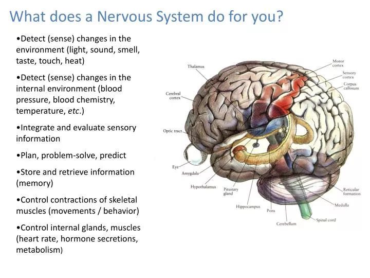
What does a Nervous System do for you?. Detect (sense) changes in the environment (light, sound, smell, taste, touch, heat) Detect (sense) changes in the internal environment (blood pressure, blood chemistry, temperature, etc .) Integrate and evaluate sensory information

E N D
What does a Nervous System do for you? • Detect (sense) changes in the environment (light, sound, smell, taste, touch, heat) • Detect (sense) changes in the internal environment (blood pressure, blood chemistry, temperature, etc.) • Integrate and evaluate sensory information • Plan, problem-solve, predict • Store and retrieve information (memory) • Control contractions of skeletal muscles (movements / behavior) • Control internal glands, muscles (heart rate, hormone secretions, metabolism)
The Nervous System Organization Central Nervous System - Brain and Spinal Cord- completely surrounded by bone - suspended in cerebrospinal fluid - covered by meninges - protected by “blood-brain barrier” Peripheral Nervous System - nerves leaving and entering the CNS - motor nerves = control muscles and glands - sensory nerves = transmit information to the CNS from sensory receptors. - cranial nerves (bottom of brain) and spinal nerves (from spinal cord)are the foundation from which all other peripheral nerves emerge.
Nervous System showing spinal nerves emerging from the spinal cord Small segment of spinal cord Showing two pairs of spinal nerves There are 31 pairs all together. Spinal Cord Functions 1. receive sensory input from receptors 2. provide motor output to muscles and glands. 3. coordinate reflexes 4. ascending and descending tracts
Quickie Review • What is included in the CNS? • What does afferent (sensory) mean and what does efferent (motor) mean? • What are cranial nerves and spinal nerves? In which division of the nervous system are they categorized? • What are three different ways that the CNS is protected?
Cells of The Nervous System Two neurons releasing neurotransmitters that act on a third neuron. The first two neurons could be in the Central Nervous System, and the third might be a motor neuron leading out to a muscle or gland. • Neuroglia (supporting cells) • Provide physical support to neurons • Provide electrical insulation to neurons • - May be involved in processes such as memory. • Neurons • Detect physical and chemical changes in their environment • Transmit electric impulses (action potentials) from one end to the other (one cell may be more than 3 feet long) • - Communicates with other neurons, muscles and glands using chemicals called neurotransmitters.
Neuroglia of the Central Nervous System
Neuroglia of the Central Nervous System Microglia - immune-like cells
Neuroglia of the Central Nervous System Oligodendrocyte produce myelin sheath
Neuroglia of the Central Nervous System Astrocyte - blood brain barrier
Neuroglia of the Central Nervous System Ependymal cells - line ventricles and help produce cerebrospinal fluid
Quickie Review • What are four different functions of the neuroglia? • The cells that function like immune cells in the CNS are the A. oligodendrocytes B. ependymal cells C. astrocytes D. microglia • The cells that form a myelin sheath around the axons of neurons are the A. oligodendrocytes B. ependymal cells C. astrocytes D. microglia
Neurons: the excitable cells of the nervous system Motor neurons have their cell bodies in the spinal cord, but their axons extend outward into the body to stimulate muscles or glands. Spinal cord smear Dendrites Microglia
Neurons: the excitable cells of the nervous system Terminal of a motor neuron axon - where the neuron stimulatesthe muscle cell to contract. Axon Axonterminus Musclecell Neuromuscular junction(motor end plate)
Occurs at the axon “hillock” If the dendrites and/or the cell body of a neuron receive sufficient stimulation, the axon Hillock will reach a threshold voltage. This voltage initiates an action potential that moves along the axon. When the action potential reaches the end of the axon, it initiates the release of neurotransmitter from the axon terminus.
Quickie Review • What are the shorter, thin extensions from a neuron cell body called? • What is the long (up to 3 feet) extension from the neuron cell body called? • What are three different types of stimuli that may effect a neuron dendrite or cell body? • What is the name of the region on a neuron where an action potential is initiated?
Action potentials are tiny electric impulses produced by neurons. They are used for transmitting information away from the cell body and toward the axon terminals. When they reach the axon terminals, the action potentials cause the release of neurotransmitter from the terminals. When a neuron is stimulated, not every stimulus will cause an action potential. The stimulus must be sufficient to cause the neuronto reach threshold. Only then will an action potential be produced.
A local depolarization at a dendrite may cause the neuron to depolarize to threshold. If that happens, an action potential will initiate at the axon hillock. dendrites Axon hillock axon Neurotransmitter bound to the gated channel. Gated channel Phospholipids
Common Traits of All Neurons • They have a polarized membrane at rest. This means that there are more positive charges on the outside than the inside. • There is a high concentration of Na+ outside the cell and a high concentration of K+ inside the cell. • The Na/K ATPase pump uses active transport to establish these concentration gradients and the membrane polarity. • During an action potential (nerve impulse), the membrane momentarily becomes “depolarized,” and then “repolarizes” back to its resting state. The processes of depolarization and repolarization involve membrane proteins called “gated channels.” • The depolarization and repolarization moves down the membrane from one end of the axon to the other.
Cell Membrane Structure + Open gated channel phospholipids Membrane glycoprotein Closedgated channel Gated channels are membrane proteins that can “open” and “close” and thereby allow the passage of ions into or out of the cell.
The Na+/K+ ATPase pump sets up conditions that make an action potential possible. K+ Three positives Pumped out Membrane protein functions as pump Only two positives Pumped in Na+ The Na/K ATPase pump establishes ion gradients for Na+ and K+ AND contributes to the polarization of the membrane (negative inside, positive outside)
The Action Potential Occurs When Na+ channels and K+ channels open sequentially Na+ channel opens, and Na+ ions rush in causingdepolarization Resting membrane potential K+ channel opens andK+ ions rush out causing repolarization
Action Potential Summary • Action potentials are not like electricity flowing through a wire. • Action potentials involve complex mechanical events at the membrane involving proteins called gated channels. • Action potentials result when Sodium (Na) and Potassium (K) ions pass through the membrane very rapidly in opposite directions. The Na+/K+ ATPase pump is NOT directlyinvolved in action potentials, but it does set up the conditions required for an AP. Action Potential Video
Quickie Review • What does it mean when we say that the resting membrane potential is “polarized?” • Which membrane protein sets up the conditions that are necessary for an action potential to occur? What are those conditions? • Which membrane proteins are actively involved during membrane depolarization? • Which membrane proteins are actively involved during membrane repolarization?
A long, straight road stretching from Indianapolis to St. Louis, MO. This represents an axon of a neuron. The white lines on each side of the road are the cell membranes of the axon. The dashed yellow line in the center of the road reminds us that the interior of the axon is negative. The crosses formed at the top of each telephone pole remind us that the extracellular environment is relatively positive.
Review of Toilet Flush-Cycle • Push on handle until it reaches threshold. • Water in bowl rises (depolarization) • Water in bowl rapidly falls (repolarization) • Water in bowl falls below resting level (hyperpolarization) • Water in bowl returns to resting level as the water in the tank refills. • Important notes: • 1. The toilet flush is an all-or-nothing event. You can’t stop it in mid-flush • 2. Some type of pumping mechanism must be present to refill the tank of the toilet and set up the conditions for the next flush. • 3. While the tank is filling, the toilet is “refractory;” that is it can’t flush again.
Now imagine an “Inspector Gadget” type of device. As the water in the bowl of the first toilet fills (reaches maximum depolarization), the gadget triggers the handle of the second toilet to reach threshold; and so the second toilet begins to flush (starts to depolarize).
But what if every toilet from Indianapolis to St. Louis had an “Inspector Gadget” device so that the second toilet caused the flushing of the third toilet . . . etc. All the way to St. Louis . . .
Summary of the Synapse • Action potential arrives at axon terminus • Calcium ions enter terminus • Calcium ions cause release of neurotransmitter • Neurotransmitter enters synapse • Some neurotransmitter binds to post-synaptic membrane receptors • Opening of post-synaptic gated channels may cause depolarization or hyperpolarization of post-synaptic membrane • Degradative enzyme may break down neurotransmitter • Neurotransmitter may be taken back up into pre-synaptic membrane and recycled.
Diseases and Treatments Relating to the Synapse • Alzheimer's Disease – progressive loss of neurons in the brain that produce acetyl choline. • Parkinson’s disease – progressive loss of neurons in the brain that produce dopamine • Some forms of depression may occur due to low levels of serotonin in some parts of the brain • Degradative enzyme inhibitors • Re-uptake pump inhibitors.