Download
1 / 65
650 likes | 917 Views
Disorders of the respiratory system. Dr. Wael H. Mansy , MD Assistant Professor College of Pharmacy King Saud University. Study objectives. Describe the general symptoms of respiratory disease .

E N D
Disorders of the respiratory system Dr. Wael H. Mansy, MD Assistant Professor College of Pharmacy King Saud University
Study objectives • Describe the general symptoms of respiratory disease. • Discuss the key features of an influenza infection. How do endemics, epidemics and pandemics differ? • Compare and contrast typical and atypical pneumonia. • List specific organisms that are associated with hospital-acquired and community-acquired pneumonia. • Discuss the possible etiology of bronchial asthma. What are some potential asthma triggers? • Compare and contrast the “early” and “late” phases of asthma in terms of their effects on the respiratory passages and clinical manifestations. • Describe how asthma attacks are classified based on frequency and severity of attacks. • Describe the various means by which asthma might be treated.
Study objectives • Compare and contrast chronic bronchitis and emphysema in terms of etiology and clinical manifestations. • Describe the different types of pneumothorax that might occur. What might cause each? • Discuss the etiology of cystic fibrosis. What are the major clinical manifestations? Why does each occur? • What is adult respiratory distress syndrome? How does it differ from respiratory distress syndrome in the newborn? • List some possible causes of interstitial lung disease. How do interstitial lung diseases differ from diseases such as emphysema and chronic bronchitis? • List some possible causes of respiratory failure. What are the major manifestations of respiratory failure?
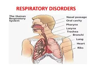
Respiratory structures such as the airways, alveoli and pleural membranes may all be affected by various disease processes. These respiratory diseases include: • Infections such as pneumonia. • Obstructive disorders that obstruct airflow into and out of the lungs such as asthma, bronchitis and emphysema. • Restrictive disorders are conditions that limit normal expansion of the lungs such as pneumothorax, atelectasis, respiratory distress syndrome and cystic fibrosis. • Cancers or exposure to Inhaled particles alter the pulmonary function.
Respiratory infections Infections of the respiratory tract can occur in: • The upper respiratory tract or • The lower respiratory tract, or • Both. Organisms capable of infecting respiratory structures include: • bacteria. • viruses: the majority of upper respiratory tract infections are caused by viruses as rhinovirus and parainfluenza virus. • fungi. Depending on the organism and extent of infection, the manifestations can range from mild to severe and even life threatening.
Upper respiratory tract Infections The common cold The most common viral pathogens for the “common cold” are rhinovirus, parainfluenza virus, respiratory syncytial virus, adenovirus and coronavirus. • These viruses tend to have seasonal variations in their peak incidence. • They gain entry to the body through the nasal mucosa and the surfaces of the eye. They are readily spread from person to person via respiratory secretions. • Manifestations of the common cold include: • Rhinitis: Inflammation of the nasal mucosa • Sinusitis :Inflammation of the sinus mucosa • Pharyngitis : Inflammation of the pharynx and throat • Headache • Nasal discharge and congestion
Upper respiratory tract Infections Influenza • Influenza is a viral infection that can affect the upper or lower respiratory tract. • Three distinct forms of influenza virus have been identified: A, B and C, of these three variants, type A is the most common and causes the most serious illness. • The influenza virus is a highly transmissible respiratory pathogen. • Because the organism has a high tendency for genetic mutation, new variant of the virus are constantly arising in different places around the world. Serious pandemics (spread of infection across a large region) of influenza are seen every 8 to 10 years as a result of this genetic mutation .
Upper respiratory tract Infections Influenza • Symptoms of influenza infection: • Headache • Fever, chills • Muscle aches • Nasal discharge • Unproductive cough • Sore throat • Influenza infection can cause marked inflammation of the respiratory epithelium leading to acute tissue damage and a loss of ciliated cells that protect the respiratory passages from other organisms. • As a result, influenza infection may lead to co-infection of the respiratory passages with bacteria. • It is also possible for the influenza virus to infect the tissues of the lung itself to cause a viral pneumonia.
Upper respiratory tract Infections Treatment of influenza: Bed rest, fluids, warmth Antiviral drugs Influenza vaccine : Provides protection against certain A and B influenza strains that are expected to be prevalent in a certain year. The vaccine must be updated and administered yearly to be effective but will not be effective against influenza strains not included in the vaccine. The influenza vaccine is particularly indicated in elderly people, in individuals weakened by other disease and in health-care workers Influenza
Lower respiratory tract Infections • The respiratory tract is protected by a number of very effective defense mechanisms designed to keep infectious organisms and particulates from reaching the lungs . • For an organism to reach the lower respiratory tract, theorganism must be particularly virulent and present in very large number or the host defense barriers must be weakened. • Factor that might weaken the respiratory defense barriers: • Cigarette smoking, which can paralyze the cilia lining the cells of the respiratory passages and impair removal of secretions, particles and microorganisms. • The presence of a respiratory pathogen such as the cold or influenza virus .
Lower respiratory tract Infections Defenses of the Respiratory System • Moist, mucus-covered surfaces : Trap particles and organisms • Cell surface IgA, lysosomes • Ciliated epithelium : Clears trapped particles and organisms from airway passages • Cough reflex and epiglottis : Prevents aspiration of particles and irritants into lower airways • Pulmonary macrophages : Phagocytize foreign particles and organisms in the alveolar spaces
Lower respiratory tract Infections Pneumonia • Pneumonia is a condition that involves inflammation of lower lung structures such as the alveoli or interstitial spaces. • It may be caused by bacteria or viruses such as pneumocystis carinii. • The prevalence and severity of pneumonia have been heightened in recent years due to the emergence of HIV as well as antibiotic resistance. • Pneumonia may be classified according to the pathogen that is responsible for the infection. • There tend to be distinct organisms that cause pneumonia in the hospital setting vs. the community setting.
Lower respiratory tract Infections Pneumonia Individuals Most at Risk for Pneumonia • Elderly • Those with viral infection • Chronically ill • AIDS or immunosuppressed patients • Smokers • Patients with chronic respiratory disease e.g. bronchial asthma.
Lower respiratory tract Infections Pneumonia Community acquired pneumonia (CAP) Aspiration pneumonia Hospital –Hospital acquired pneumonia (HAP) –Ventilator associated pneumonia (VAP) –Healthcare associated pneumonia (HCAP)
Potential Pathogens Bacteria *Typical Streptococcus pneumoniae Hemophilusinfluenzae Mycobacterium catarrhalis Klebsiellapneumoniae *Atypical Chlamydiapneumoniae Legionellapneumophila Mycoplasmapneumoniae.
Viruses Fungi Less Common pathogens – N. meningitidis – Chlamydia psittaci – B. anthracis – Y. pestis
Lower respiratory tract Infections Pneumonia A second classification scheme for pneumonia is based on the specific structures of the lung that the organisms infect and includes typical and atypical pneumonia. Typical pneumonia • Usually bacterial in origin. • Organisms replicate in the spaces of the alveoli. Manifestations: • Inflammation and fluid accumulation are seen in the alveoli. • White cell infiltration and exudation can been seen on chest radiographs. • High fever, chest pain, chills, and malaise are present. • Purulent sputum is present. • Some degree of hypoxemia is present.
Lower respiratory tract Infections Pneumonia Atypical pneumonia • Usually viral in origin. • Organisms replicate in the spaces around the alveoli. Manifestations: • Milder symptoms than typical pneumonia. • Lack of white cell infiltration in alveoli. • Lack of fluid accumulation in the alveoli. • Not usually evident on radiographs. • May make the patient susceptible to bacterial pneumonia.
Lower respiratory tract Infections Pneumonia Opportunistic organisms • A number of organisms not commonly associated with respiratory illness in otherwise healthy individuals can cause severe respiratory infections and pneumonia in patients with HIV or those who are immunocompromised as a result of immune suppressive therapy. • These organisms include mycobacteria, fungus (Histoplasma) and protozoa (Pneumocystis carinii). • Treatment of these organisms requires specific drug therapy, and, in the case of protozoa and fungi, the organisms are very difficult to kill.
Lower respiratory tract Infections Pneumonia Treatment of pneumonia: • Antibiotics if bacterial in origin. The health-care provider should consider the possibility that antibiotic-resistant organisms are present. • Oxygen therapy for hypoxemia. • A vaccine for pneumococcal pneumonia is currently available and highly effective. This vaccine should be considered in high-risk individuals.
Bronchial asthma Asthma is a chronic inflammatory disorder of the airways in which many cells and cellular elements play a role: in particular, mast cells, eosinophils, neutrophils, T lymphocytes, macrophages, and epithelial cells. In susceptible individuals, this inflammation causes recurrent episodes of coughing (particularly at night or early in the morning), wheezing, breathlessness, and chest tightness. These episodes are often reversible either spontaneously or with treatment. A key component of asthma appears to be airway “hyper reactivity” in affected individuals. Exposure to certain “triggers” can induce marked bronchospasm and airway inflammation in susceptible patients
Obstructive Respiratory Disorders Bronchial asthma • Individuals with asthma appear to produce large amounts of the antibody IgE that attach to the mast cells present in many tissues. • Exposure to a trigger such as pollen will result in the allergen-binding mast cell-bound IgE, which in turn causes the release of inflammatory mediators such as Histamine , Leukotrienes and Eosinophilic Chemotactic factor. • The response of a patient with asthma to these triggers can be divided into an “early phase” and a “late phase.”
Obstructive Respiratory Disorders Bronchial asthma **Some Potential Asthma Triggers** • Allergens — Pollen, pet dander, fungi, dust mites • Cold air • Pollutants • Cigarette smoke • Strong emotions • Exercise • Respiratory tract infections
Bronchial asthma Intrinsic (Nonatopic) Asthma. Intrinsic or nonatopic asthma triggers include: respiratory tract infections, exercise, hyperventilation, cold air, exercise, drugs and chemicals, hormonal changes and emotional upsets, airborne pollutants, and gastroesophageal reflux.
Bronchial asthma Extrinsic or atopic asthma is typically initiated by a type I hypersensitivity reaction and is seen in persons with a family history of atopic allergy. Person with atopic asthma often have other allergic disorders, such as hay fever, urticaria, and eczema. Among airborne allergens implicated in atopic asthma are house dust mite allergens, cockroach allergens, animal danders.
Clinical course, severity Daytime asthma symptoms Nighttime awakenings FEV1, PEF < 1 /week 2 and < /month >80% predicted. Daily variability < 20% Mild Intermittent Mild persistent 1 /week but not daily > 2 /month >80% predicted. Daily variability – 20-30% Daily are severe enough to affect activity Moderate persistent > 1 /week > 60 but < 80% predicted. Variability>30%. Severe persistent Persistent, which limit normal activity Daily <60% predicted. Variability > 30%. Asthma severity classification
Bronchial asthma During an asthmatic attack, the airways narrow because of bronchospasm, edema of the bronchial mucosa, and mucus plugging. Expiration becomes prolonged because of progressive airway obstruction. The amount of air that can be forcibly expired in 1 second (forced expiratory volume in 1 second [FEV1.0]) and the peak expiratory flow rate (PEF), measured in liters per second, are decreased
Obstructive Respiratory Disorders Bronchial asthma Early phase of asthma: The early phase of asthma is characterized by: • marked constriction of bronchial airways (bronchospasm) • edema of the airways • production of excess mucus. The bronchospasm that occurs may be the result of the increased release of certain inflammatory mediators such as histamine, prostaglandins and bradykinin that, in the early stages of asthmatic response, promote bronchoconstriction rather than inflammation.
Obstructive Respiratory Disorders Bronchial asthma Late phase of asthma: • The late phase of asthma can occur several hours after the initial onset of symptoms and manifests mainly as an inflammatory response. • The primary mediators of inflammation during the asthmatic response are the white blood cells Eosinophilsthat stimulate mast cell degranulation and release substances that attract other white cells to the area. • Subsequent infiltration of the airway tissues with white blood cells such as Neutrophils and lymphocytes also contributes to the overall inflammatory response of the late phase of asthma.
Obstructive Respiratory Disorders Bronchial asthma Manifestations of asthma : • Coughing, wheezing • Difficulty breathing • Rapid, shallow breathing • Increased respiratory rate • Excess mucus production • Significant anxiety
Obstructive Respiratory Disorders Bronchial asthma Staging of the Severity of an Acute Asthma Attack • Stage I (mild) • Mild Dyspnea • Diffuse wheezing • Adequate air exchange • Stage II (moderate) • Respiratory distress at rest • Marked wheezing • Stage III (severe) • Marked respiratory distress • Cyanosis • Marked wheezing or absence of breath sounds • Stage IV (respiratory failure) • Severe respiratory distress, lethargy and confusion
Obstructive Respiratory Disorders Bronchial asthma Possible complications of asthma can include : • Severe acute Asthma (status asthmatics), which is a life-threatening condition of prolonged bronchospasm that is often not responsive to drug therapy. • Pneumothorax : is also a possible consequence as a result of lung pressure increases that can result from the extreme difficulty involved in expiration during a prolonged asthma attack. • Respiratory failure: marked hypoxemia and acidosis might occur.
Obstructive Respiratory Disorders Bronchial asthma Treatment of asthma: The appropriate drug treatment regimen for asthma is based on the frequency and severity of the asthma attacks and may include the following: • 1. Avoidance of triggers, and allergens. Improved ventilation of the living spaces, use of air conditioning. • 2. Bronchodilators(examples: albuterol, terbutaline): Short acting β-Adrenergic receptor activators. May be administered as needed in the form of a nebulizer solution using a metered dispenser or may be given subcutaneously. These drugs block bronchoconstriction but do not prevent the inflammatory response.
Obstructive Respiratory Disorders Bronchial asthma Treatment of asthma: 3.Xanthine drugs (example: theophylline) : • Cause bronchodilation and also inhibit the late phase of asthma. • These drugs are often used orally as second-line agents in combination with other asthma therapies such as steroids. • Drug like theophylline can have significant central nervous system, cardiovascular and gastrointestinal side effects that limit their overall usefulness. 4.Cromolyn sodium : Anti-inflammatory agent that blocks both the early and late phase of asthma. The mechanism of action is unclear but may involve mast cell function or responsiveness to allergens.
Obstructive Respiratory Disorders Bronchial asthma Treatment of asthma: • 5. Anti-inflammatory drugs (corticosteroids) : Used orally or by inhalation to blunt the inflammatory response of asthma. The most significant unwanted effects occur with long-term oral use of corticosteroids and may include immunosuppression , increased susceptibility to infection, osteoporosis and effects on other hormones such as the glucocorticoids. • 6. Leukotrienes modifiers (example: Zafirlukast) : New class of agents that blocks the synthesis of the key inflammatory mediators, leukotrienes.
Obstructive Respiratory Disorders Bronchitis • Bronchitis is an obstructive respiratory disease that may occur in both acute and chronic forms. • Acute bronchitis: Inflammation of the bronchial passages most commonly caused by infection with bacteria or viruses. • Acute bronchitis is generally a self-limiting condition in healthy individuals but can have much more severe consequences in individuals who are weakened with other illness or who are immunocompromised. • Symptoms of acute bronchitis often include productive cough, Dyspnea and possible fever.
COPD=Chronic Obstructive Pulmonary Disease COPD is characterized by chronic and recurrent obstruction of airflow. Airflow obstruction usually is progressive and is accompanied by inflammatory responses to noxious particles or gases. The term COPDencompasses two types of obstructive airway disease: emphysema and chronic obstructive bronchitis. Persons with COPD often have overlapping features of both disorders.
Obstructive Respiratory Disorders Chronic Bronchitis • Chronic bronchitis: Chronic bronchitis is a chronic obstructive pulmonary disease that is most frequently associated with cigarette smoking (approximately 90% of cases). • Chronic bronchitis may also be caused by prolonged exposure to inhaled particulates such as coal dust or other pollutants. • The disease is characterized by excess mucus production in the lower respiratory tract. This mucus accumulation can impair function of the ciliated epithelium and lining of the respiratory tract and prevent the clearing of debris and organisms. As a result, patients with chronic bronchitis often suffer repeated bouts of acute respiratory infection. • Chronic bronchitis sufferers are often referred to as “blue bloaters” as a result of the cyanosis and peripheral edema that is often present.
Obstructive Respiratory Disorders Chronic Bronchitis Manifestations of chronic bronchitis: • Productive, chronic cough • Production of purulent sputum • Frequent acute respiratory infections • Dyspnea • Hypoxia, cyanosis • Symptoms of cor pulmonale • Fluid accumulation (edema) in later stages
Obstructive Respiratory Disorders Chronic Bronchitis Treatment of chronic bronchitis: 1. Cessation of smoking or exposure to irritants 2. Bronchodilators to open airway passages 3. Expectorants to loosen mucus 4. Anti - inflammatory to relieve airway inflammation and reduce mucus secretion 5. Prophylactic antibiotics for respiratory infections 6. Oxygen therapy
Obstructive Respiratory Disorders Emphysema Alveolar duct Respiratory bronchioles Terminal bronchiole septum alveoli NORMAL ACINUS • Emphysema is a respiratory disease that is characterized by destruction and permanent enlargement of terminal bronchioles and alveolar air sacs
Obstructive Respiratory Disorders Emphysema • Well over 95% of all patients with emphysema were chronic cigarette smokers. Although the exact etiology of emphysema is still uncertain, • Chronic exposure to cigarette smoke causes chronic inflammation of the alveolar airways, which results in infiltration by lymphocytes and macrophages. • Excess release of protease enzymes such as trypsin from lung tissues and leukocytes can digest and destroy the elastic walls of the alveoli. • Levels of a protective enzyme α-1-antitrypsin have been shown to be lacking in certain individuals who are chronic cigarette smokers. This enzyme inactivates destructive protease enzymes (trypsin)in lung tissue. • In fact, a rare form of emphysema occurs in individuals who are not cigarette smokers but who have a genetic lack of α-1-antitrypsin.
Obstructive Respiratory Disorders Emphysema Mainly caused by: Loss of alveolar (lung) elasticity and a decrease in the overall surface area for gas exchange within the lungs. Manifestations include the following: • Tachypnea (increased respiratory rate): Because that is effective in maintaining arterial blood gases, one does not usually see hypoxia or cyanosis until the end stages of the disease. • Barrel chest from prolonged expiration. • Lack of purulent sputum. • Possible long-term consequences, including cor pulmonale , respiratory failure.
Obstructive Respiratory Disorders Emphysema Comparison of Symptoms for Chronic Bronchitis and Emphysema
Restrictive Pulmonary Disorders Pneumothorax • Pneumothorax is the entry of air into the pleural cavity in which the lungs reside. • In order for normal lung expansion to occur, there must be a negative pressure within the pleural cavity with respect to atmospheric pressure outside the pleural cavity. The inside of the pleural cavity is essentially a vacuum and when air enters the pleural cavity the negative pressure is lost and the lungs collapse. • Because each lung sits in a separate pleural cavity, pneumothorax of one plural cavity will not cause collapse of the other lung.
Restrictive Pulmonary Disorders Pneumothorax Types of pneumothorax: 1. Open or communicating pneumothorax • Usually involves a traumatic chest wound. • Air enters the pleural cavity from the atmosphere. • The lung collapses due to equilibration of pressure within the pleural cavity with atmospheric pressure. • 2. Closed or spontaneous pneumothorax • • Occurs when air “leaks” from the lungs into the pleural cavity. • • May be caused by lung cancer, rupture, pulmonary disease. • • The increased plural pressure prevents lung expansion during inspiration and the lung remains collapsed.
Restrictive Pulmonary Disorders Pneumothorax 3. Tension pneumothorax • A condition in which there is a one-way movement of air into but not out of the pleural cavity. • May involve a hole or wound to the pleural cavity that allows air to enter and the lung to collapse. Upon expiration, the hole or opening closes, which prevents the movement of air back out of the pleural cavity. • A life-threatening condition because pressure in the pleural cavity continues to increase and may result in further lung compression or compression of large blood vessels in the thorax or the heart.
Restrictive Pulmonary Disorders Pneumothorax • Manifestations of pneumothorax: • Tachypnea, Dyspnea • Chest pain • Possible compression of thoracic blood vessels and heart, especially with tension pneumothorax • Treatment of pneumothorax: • Removal of air from the pleural cavity with a needle or chest tube • Repair of trauma and closure of opening into pleural cavity