Download
1 / 32
830 likes | 1.83k Views
Imaging Anatomy of the Pancreas. Ben Barnard Department of Diagnostic Radiology; KHC. Outline. Embryology Variants Gross anatomy Anatomical relations Arterial supply Venous drainage Lymphatic drainage Radiological anatomy. Embryology.

E N D
Imaging Anatomy of the Pancreas Ben Barnard Department of Diagnostic Radiology; KHC
Outline • Embryology • Variants • Gross anatomy • Anatomical relations • Arterial supply • Venous drainage • Lymphatic drainage • Radiological anatomy
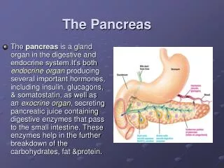
Embryology • The pancreas is formed by dorsal and ventral buds originating from the endodermal lining of the duodenum • The smaller ventral bud arises in conjunction with the bile duct and consists of two portions • Left bud which atrophies during development • Right bud which swings posteriorly and unites with the inferior division of the dorsal bud
Variants • Agenesis of the dorsal pancreatic moeity • Pancreas with a head but no body or tail – very rare • Left-sided pancreas • Effect of age with laxity of suspensory fascia • Accessory nodules (pancreatic rests) • Can occur in the wall of the stomach, duodenum, jejunum , appendix, Meckel’sdiverticulum (1-15% ) • Pancreas divisum • Failure of fusion of dorsal and ventral moeities • Anterosuperior head and body drain via accessory ampulla • Posterioinferior head drains to ampulla • Annular pancreas
Annular pancreas • Occurs when the left portion of the ventral bud fails to atrophy and migrates around the duodenum in the opposite direction to the normal right portion • Duodenum is thus surrounded bypancreatic tissue, and an annular pancreas is formed • This tissue constricts the duodenum and may cause complete obstruction
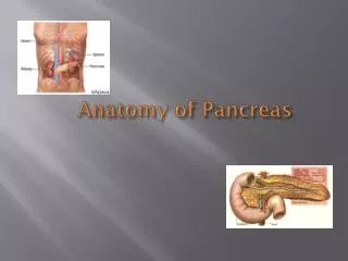
Gross Anatomy • Retroperitoneal organ • 15cm long • Transverse mesocolon originates from anterior surface.
Head • C-curve of duodenum (L2) • Uncinate process • Inferior-medial extension of head under SMA (L2) • Neck • Overlies L1 vertebral body immediately to the left of the head • Body • Extends left and slightly superior (L1) • Tail • Tapering portion extending to hilum of spleen (T12)
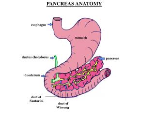
Anatomical Relations • Anterior relations • Peritonium • 1st part duodenum • Lesser sac and stomach • Transverse colon/mesocolon
Anatomical Relations • Posterior relations • CBD • IVC • PV • Aorta • SMA • SMV • Right renal vein • Left kidney • Cruraeof diaphragm.
Retroperitoneal Compartments • The pancreas (P) lies in the anterior pararenal space (APRS) together with the ascending and descending colon (C) and duodenum • ST = stomach • S = spleen • RK = right kidney • LK = left kidney • A = aorta • V = inferior vena cava • LS = lesser sac • PRS = perirenal space • PPRS = posterior pararenal space
Pancreatic duct • Pancreatic duct measures • 3mm in the head • 2mm in the neck • 1mm in the tail • Unites with the common bile duct to form the ampullaof Vater and opens into the 2ndpart of duodenum
Pancreatic duct variants • Santorini • Wirsung
Arterial supply • Pancreas neck, body and tail • Dorsal pancreatic artery (from splenic artery or coeliactrunk) • Pancreaticamagna artery ( emerges halfway along the splenic artery) • Transverse pancreatic artery (travels along the length of the pancreatic duct and anastomoseswith the above arteries) • Pancreas head • Superior pancreaticoduodenal artery (anterior and posterior branches from the gastroduodenal artery) • Inferior pancreaticoduodenal artery (anterior and posterior branches from the superior mesenteric artery)
Venous drainage • SMV • Splenic vein • Portal vein
Lymphatic drainage • Along arterial blood supply to pre-aortic coeliac lymph nodes
US • 3 - 7.5 MHz probe • Sagittal, transverse and oblique planes • Occasionally, may have non-visualization due to bowel gas which can be overcome by: • Fill stomach with fluid • Position patient erect (liver descends) • Prone position for tail
CT • Similar density as liver • Density is reduced with increasing age due to normal accumulation of fat • Homogenous enhancement post contrast
References • Imaging Anatomy of the Pancreas Referaat, Dr FransNaude • Normal Findings in CT and MRI,Moeller • Pocket Atlas of Sectional Anatomy Computer Tomography and Magnetic Resonance Imaging, Torsten B. Moeller, MD • Last’s anatomy 11thed , Sinnatamby • Applied radiological anatomy , Butler • Atlas of human anatomy, F Netter • Pocket Atlas of Radiographic Anatomy,secondedition,Moeller • Atlas of normal rontgen variants that may simulate disease, R Keats • Color Atlas of Ultrasound Anatomy ,Berthold Block • Grant atlas of anatomy