Download
1 / 30
920 likes | 2.19k Views
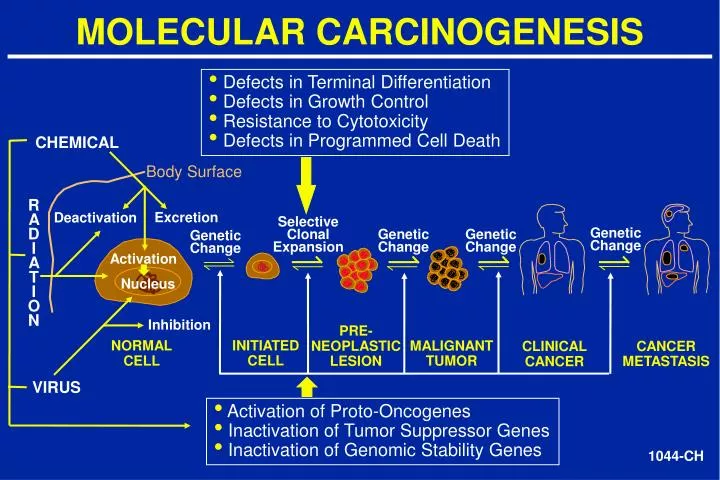
MOLECULAR CARCINOGENESIS. Defects in Terminal Differentiation Defects in Growth Control Resistance to Cytotoxicity Defects in Programmed Cell Death. CHEMICAL. Body Surface. R A D I A T I O N. Excretion. Deactivation. Selective Clonal Expansion. Genetic Change. Genetic

E N D
MOLECULAR CARCINOGENESIS • Defects in Terminal Differentiation • Defects in Growth Control • Resistance to Cytotoxicity • Defects in Programmed Cell Death CHEMICAL Body Surface R A D I A T I O N Excretion Deactivation Selective Clonal Expansion Genetic Change Genetic Change Genetic Change Genetic Change Activation Nucleus Inhibition PRE- NEOPLASTIC LESION INITIATED CELL NORMAL CELL MALIGNANT TUMOR CANCER METASTASIS CLINICAL CANCER VIRUS • Activation of Proto-Oncogenes • Inactivation of Tumor Suppressor Genes • Inactivation of Genomic Stability Genes 1044-CH
CHRONIC INFLAMMATION AND CANCER Inherited Acquired Disease Tumor Site Risk Hemochromatosis Liver 219 Hereditary Pancreatitis Pancreas 120 Crohn’s Disease Colon 3 Ulcerative Colitis Colon 6 Disease Tumor Site Risk Viral Hepatitis B Liver 88 Hepatitis C Liver 3 Bacterial Helicobacter Pylori Gastric 11 PID Ovary 3 Parasitic S. hematobium Urinary Bladder 2-14 S. japonicum Colon 2-6 Liver Fluke Liver 14 Chemical/ Physical Acid reflex Esophagus 50-100 Metabolic Disease Obesity Colon 1.5 “Chronic infection and associated inflammation contribute to about 1/3 of cancers worldwide” -B.N. Ames, PNAS, 1995 “18% of human cancers, i.e., 1.6 million per year, are related to infection.” - B. Stewart and P. Kleihues World Cancer Report, IARC Press, p. 57, 2003
CANCERS ASSOCIATED WITH OBESITY In Women In Men • Breast (postmenopausal) • Endometrium • Cervical • Ovarian • Colorectal • Kidney • Liver/ Gall Bladder • Pancreatic • Esophageal • Hematopoietic • Prostate • Colorectal • Kidney • Liver/Gall Bladder • Pancreatic • Esophageal • Hematopoietic Calle, E et al., NEJM 348:1625-38, 2003 3000-CH
REACTIVE NITROGEN AND OXYGEN SPECIES DERIVED FROM INFLAMMATORY CELLS HOCl HOBr Oxidation & Halogenation H2O2 +Cl-/Br- Myeloperoxidase NO2• NO2- Oxidation & Nitration O2 Neutrophil N2O3 Deamination Nitrous Anhydride NO• - OH • iNOS ONOO NO2• Oxidation & Nitration - CO2 O2• CO3• SOD ONOOCO2- Of DNA and Proteins Macrophage Nitrosoperoxycarbonate H2O2 2786*-CH
FREE RADIALS AND INFLAMMATION ROS •OH O2- • (Hydroxyl (Superoxide) radical) RNS NO • ONOO- N2O3 (Nitric Oxide) (Peroxynitrite) (Nitroxyl Radical) Protein Damage (DNA Repair Enzymes, Caspases) Lipid Peroxidation DNA Damage and Mutation MDA (malondialdehyde) 4HNE (4-hydroxynonenal) Arachidonic Acid Cascade Nitrosamines/Deamination 8--oxo-dG 8-nitroguanine Etheno Adducts M1G Adduct S-nitrosothiol SSB’s DSB’s Eicosenoids Cell Proliferation 1760-CH
CaM CaM CaM CaM CaM CaM NITRIC OXIDE SYNTHASE Inactive cNOS Inactive cNOS Active cNOS Ca+++2 NO Citrulline L-ARGININE NO iNOS Always active Billiar
NITRIC OXIDE DAMAGES DNA AND ACTIVATES p53 IN MCF-7 CELLS p53 MODIFICATIONS DNA DAMAGE SPER/NO p53 P-Ser-15 MCF-7 Cells P-Ser-20 P-Ser-33 P-Ser-46 P-Ser-315 P-Ser-392 K-Lys-382 2016*-CH
NO-INDUCED p53 PHOSPHORYLATION TRANSACTIVATES DOWNSTREAM PROTEINS AND ENGAGES A G2/M ARREST % Cells in G2/M DOWNSTREAM PROTEINS Mitotic Index 2122A*-CH
INDUCIBLE NITRIC OXIDE SYNTHASE (NOS2)AND CYCLOOXYGENASE-2 (COX2)INTERACTIONS IN HUMAN CARCINOGENESIS Genomic instability HIF1a Hypoxia NOS2 p53 • Selective Clonal Expansion • DNA damage Mutant p53 • NO Cytokines e.g., IL-1b TNF-a Lipid Peroxidation Hypoxia COX2 Apoptosis Prostaglandins (e.g., PGE2) NFB p53 K-ras 1079A*-CH
VENN DIAGRAM OF 1396 “p53-DEPENDENT” GENES MODULATED BY CELLULAR STRESS NO H2O2 HU Hypoxia 29 139 genes 666 genes 34 35 14 33 40 5 14 11 4 225 genes 140 genes 7 HU Hypoxia (T-test at p<0.001 for each treatment and time point) 2978-CH
EXAMPLES OF CHRONIC INFLAMMATORY CONDITIONS ASSOCIATED WITH INCREASED p53 MUTATION LOAD • ULCERATIVE COLITIS • HEMOCHROMATOSIS • WILSON DISEASE • VIRAL HEPATITIS
-7 Absolute Mutation frequency x 10 p53 MUTATION LOAD IS INCREASED IN ULCERATIVE COLITIS 30 G TO A (CpG SITE OF CODON 248 ) UC vs. Non-UC (p < 0.001) C TO T (CODON 247) UC vs. Non-UC ( p < 0.001) 2 0 1 0 0 ULCERATIVE NORMALCONTROL COLITIS 054-PH
Tumor Suppressor (1990s) Transcription Cell Cycle Checkpoints Senescence Protooncogene (1980s) p53 (1979) Programmed Cell Death Development • DNA Repair • Homologous Recombination • Chromosomal Segregation Genomic Stability (1990s) 509B*-CH Ageing (2000s)
p53 IS AT THE CROSSROADS OF CELLULAR STRESS RESPONSE PATHWAYS DNA Damage Hypoxia Oncogene Activation E2F NOS2 p14ARF ATM, ATR, CHK2 p53 mdm2 Cell Cycle Checkpoints Apoptosis DNA Repair Senescence p21WA F1 14-3-3sGadd45 p21WAF1 Others PUMA, NOXA, BAX, Apaf1, XPB, XPD,WRN, BLM GADD45, p48, p53R2 APE1, Pol b 1306I*-CH
EXAMPLES OF p53 NEGATIVE FEEDBACK LOOPS • Posttranslational Modification and Proteolytic Cleavage Oliver et al., Nature 362: 857, 1993 Wu et al., Genes Dev. 7: 1126, 1993 • ATM-Dependent DNA Damage Pathway Matsui et al., J. Biol. Chem. in press, 2004 • Nitric Oxide Pathway p53 MDM2 Ubiquitination CHK2 Kinase p53 Phosphorylation Transrepression Inducible Nitric Oxide Synthase DNA Damage Transrepression p53 Forrester et al., PNAS 93: 2442, 1996 Ambs et al., PNAS 95: 8823, 1998 2900-CH
MODEL OF CELLULAR STRESS INDUCED p53 ACTIVATION AND APOPTOSIS Cellular Stress MnSOD GPX1 Mitochondrial Depolarization • H2O + O2 H2O2 O2 CAT Fe2+ PUMA, NOXA, BAX, p53AIP1 PIG3 Ferredoxin reductase p53 Lipid Peroxidation Cytochrome C + ProCaspase 9 + APAF1 APOPTOSOME APOPTOSIS EXECUTIONER CASPASES 1996A*-CH
APAF-1 IS A TRANSCRIPTIONAL TARGET OF p53 IN DNA DAMAGE-INDUCED APOPTOSIS p53 Apoptotic stimuli RE PUMA, NOXA, p53AIP1, Bax Mitochondrial depolarization Apoptosome Pro-caspase 9 Apaf-1 Cytochrome C Caspase 3 APOPTOSIS 1917*-CH
CELL DEATH Non-programmed Programmed 1935-CH
MUTATIONAL SPECTRA OF THE p53, APC, ATM AND BRCA-1 GENES IN ALL HUMAN CANCERS p53 (n=15,122) APC (n=1,451) Nonsense 7% Frameshift 9% Nonsense 32% Splice site 2% In Frame Del/Ins. 2% Frameshift 51% Silent 5% Missense 75% Missense 4% Silent 9% Splice site 4% ATM (n=617) BRCA-1 (n=3,703) Nonsense 11% Nonsense 14% Frameshift 56% Frameshift 54% Missense 30% Missense 28% Splice site 5% In Frame Del/Ins. 2% 797B-CH
Sunlight Skin, Codon 281 Aflatoxin B1 and HBV Liver, Codon 249 G:C to T:A 98% CC to TT 100% EXAMPLES OF p53 MUTATION HOTSPOTS ASSOCIATED WITH CARCINOGEN EXPOSURE Hemochromatosis Liver, Codon 220 Tobacco Smoking Lung ,Codon 157 400 249 300 200 G:C to T:A 78% 281 A:T to G:C 100% 220 157 Missense 100 0 97 292 324 352 17-29 EVOLUTIONARILY CONSERVED N C Sequence-Specific DNA Binding Domain Oligomerization and Nuclear Localization and Export Domains Transactivation Domain 567L-CH
HYPOTHESIS: • p53 mutation hotspots in clonally derived human cancers reflect the preferential: • sites of carcinogen-DNA adduct formation in the gene • sites of slow repair of DNA damage • mutagenic potential of certain carcinogen-DNA adducts • pathobiological effects of the p53 mutant leading to a selective clonal expansion advantage, including “gain of function” or an increase in genomic instability 1156-CH
WORLDWIDE p53 MUTATIONAL SPECTRA IN HCC FROM DIFFERENT GEOGRAPHICAL AREAS < 3.3 p53 MUTATION DIAGRAM < 5.6 Del + ins. G:C to C:G < 9.0 A:T to C:G G:C to T:A < 15.0 G:C to A:T A:T to G:C CpG < 98.9 G:C to A:T A:T to T:A Non-CpG Japan N=242 North America N=15 Western Europe N=82 China N=171 Taiwan N=113 Africa N=28 Incidence of HCC per 100,000 2115-CH
ASSESSMENT OF CAUSATION BY THE BRADFORD-HILL CRITERIA HYPOTHESIS: Dietary aflatoxin B1 exposure can produce 249ser (AGG->AGT) p53 mutations during human liver carcinogenesis • STRENGTH OF ASSOCIATION • Consistency • Positive correlation in 3 different ethnic populations on 3 continents • Temporality • 249ser p53 mutant cells observed in non-tumorous liver in high HCC incidence geographic areas • Specificity • 249ser p53 mutations are uncommon in other cancer types • 249ser p53 mutation in serum DNA is a biomarker of liver cancer risk 926C-CH From: Hussain and Harris, Cancer Res. 58: 4023-37, 1998
ASSESSMENT OF CAUSATION BY THE BRADFORD-HILL CRITERIA HYPOTHESIS: Dietary aflatoxin B1 exposure can produce 249ser (AGG->AGT) p53 mutations during human liver carcinogenesis • BIOLOGIC PLAUSIBILITY • AFB1 is a potent carcinogen in rodents, monkeys and humans • AFB1 is enzymatically activated by human hepatocytes to 8,9-AFB1 oxide that binds to DNA, including the 3rd base (G) at codon 249 • AFB1 exposure to human liver cells in vitro produces codon 249ser p53 mutations • 249ser p53 expression inhibits apoptosis and p53-mediated transcription and enhances liver cell growth rates in vitro From: Hussain and Harris, Cancer Res. 58: 4023-37, 1998 926D-CH
p53 CODON 249ser MUTANT IN SERUM DNA AND SERUM HBVSAg ARE BIOMARKERS OF LIVER CANCER RISK • HBSAg/249p53 mutantRR(95%CI) minus/minus 1 plus/minus 10(5-20) minus/plus 13(5-35) plus/plus 399(49-3272) Kirk, GD et al., Proc. 11th Int. Symposium on Viral Hepatitis and Liver Diseases, Sydney, 2003. FORTY PERCENT OF LIVER CANCER IN QIDONG, PRC IS ATTRIBUTABLE TO AFLATOXIN DIETARY EXPOSURE Ming, l et al., Hepatology 36: 1214-20, 2002.
COLORECTAL CARCINOGENESIS • SPORADIC: Normal Adenomatous Mucosa Polyps Carcinoma • ULCERATIVE COLITIS ASSOCIATED: Ulcerative Colitis Dysplasia Carcinoma • Mutation • K-ras APC -catenin p53 • ~45% 85% ~10% ~55% • ~15% 6% ?% ~55% 885-CH
EXAMPLES OF GENETIC LESIONSIN BRONCHIAL DYSPLASIA, CARCINOMA-IN-SITU AND LUNG CARCINOMA Lesion Dysplasia CIS Carcinoma • LOH • 3p12, 14, 21 • 9p21 • 17p13 • p53 (p17p13) • p16 (9p21) • Telomerase • Ki-ras • FHIT (3p14) • Rb 1049-CH