Download
1 / 27
300 likes | 368 Views
Cardiac Cycle. Blood Flow Through Heart. Definitions. Systole = period of ventricular contraction. Diastole = period of ventricular relaxation. NOTE: Normally diastole is longer than systole. Cardiac cycle. General Principles.

E N D
Definitions • Systole = period of ventricular contraction. • Diastole = period of ventricular relaxation. • NOTE: Normally diastole is longer than systole.
Cardiac cycle • General Principles. • Contraction of the myocardium generates pressure changes which result in the orderly movement of blood. • Blood flows from an area of high pressure to an area of low pressure, unless flow is blocked by a valve. • Events on the right and left sides of the heart are the same, but pressures are lower on the right.
Atrial systole • The heart is full of blood and the ventricles are relaxed • Both the atria contract and blood passes down to the ventricles • The atrio-ventricular valves open due to blood pressure • 70% of the blood flows passively down to the ventricles so the atria do not have to contract a great amount.
Ventricular systole • The atria relax. • The ventricle walls contract, forcing the blood out • The pressure of the blood forces the atrio-ventricular valves to shut (producing the heart sound ‘lub’)
Ventricular systole • The pressure of blood opens the semi-lunar valves. • Blood passes into the aorta and pulmonary arteries.
Diastole • The ventricles relax • Pressure in the ventricles falls below that in the arteries • Blood under high pressure in the arteries causes the semi lunar valves to shut. This produces the second heart sound, ‘dub’. • During diastole, all the muscle in the heart relaxes.
Blood from the vena cava and pulmonary veins enter the atria. • The whole cycle starts again.
CARDIAC CYCLE • The normal duration of the cardiac cycle is 0.8 second. • Each beat of the heart consists of systole and diastole of atria and ventricles. • Systole is the contraction of the heart during which blood is ejected out from the heart • Diastole is the relaxation of the heart during which the chambers of the heart are filled with blood
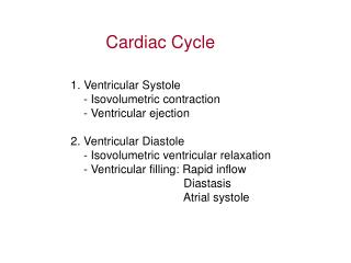
Ventricular Systole: 1. Isovolumetric contraction phase - 0.05 sec 2. Rapid Ejection phase - 0.10 sec 3. Reduced Ejection phase - 0.15 sec
Ventricular diastole • Proto diastolic phase - 0.04 sec • Isovolumetric Relaxation phase - 0.06 sec • First Rapid filling phase - 0.10 sec • Slow Filling - 0.20 sec • Last Rapid Filling Phase - 0.10 sec
FACTS TO REMEMBER • SYSTOLE IS CONTRACTION -THE RISE IN PRESSURE IN THE CONTRACTING CHAMBER -BLOOD BEING EJECTED BY THE CONTRACTING CHAMBER • DIASTOLE IS RELAXATION - A FALL IN PRESSURE OF THE RELAXING CHAMBER - FILLING OF THE RELAXING CHAMBER
ATRIAL SYSTOLE - Heart • Prior to atrial systole, blood has been flowing passively from the atrium into the ventricle through the open AV valve. • Contraction of atria propels some additional blood into the ventricles. Atrial contraction is complete before the ventricle begins to contract.
ISOVOLUMETRIC CONTRACTIONHeart • The atrioventricular (AV) valves close at the beginning of this phase. • Electrically, ventricular systole is defined as the interval between the QRS complex and the end of the T wave (the Q-T interval). • Mechanically, ventricular systole is defined as the interval between the closing of the AV valves and the opening of the semilunar valves (aortic and pulmonary valves).
RAPID EJECTIONHeart • The semilunar (aortic and pulmonary) valves open at the beginning of this phase.
REDUCED EJECTIONHeart • At the end of this phase the semilunar (aortic and pulmonary) valves close.
ISOVOLUMETRIC RELAXATIONHeart • At the beginning of this phase the AV valves are closed.
RAPID VENTRICULAR FILLINGHeart • Once the AV valves open, blood that has accumulated in the atria flows rapidly into the ventricles.
REDUCED VENTRICULAR FILLINGHeart • Rest of blood that has accumulated in the atria flows slowly into the ventricles.
Heart sounds When the valves close, the surrounding fluids vibrate under the influenceof sudden pressure changes, giving off sound that travels in all directions through the chest. When the ventricles contract, one first hears a sound caused by closure of the A-V valves. The vibration is low in pitch and relatively long-lasting and is known as the first heart sound. When the aortic and pulmonary valves close at the end of systole, one hears a rapid snap because these valves close rapidly, and the surroundings vibrate for a short period. This sound is called the second heart sound.
First heart sound : It is due to the closure of AV valves ( mitral and tricuspid valves) . Heard as LUB Duration is 0.09 to 0.15 seconds It is characterized by prolonged ,loud sound and best heard at mitral and tricuspid areas
Second heart sound: : • It is due to the closure of semilunar valves. Heard as DUP, Duration is 0.10 seconds It is characterized by short,sharp sound and best heard at aortic and pulmonary areas.