Download
1 / 1
10 likes | 148 Views
Development of a compact gamma camera for intra operative radiation imaging Konstantinou G. 1 , Chil R. 1 , Desco M. 1,2 , and Vaquero J.J. 1,2 1 Departamento de Bioingeniería e Ingeniería Aeroespacial, Universidad Carlos III de Madrid. Spain

E N D
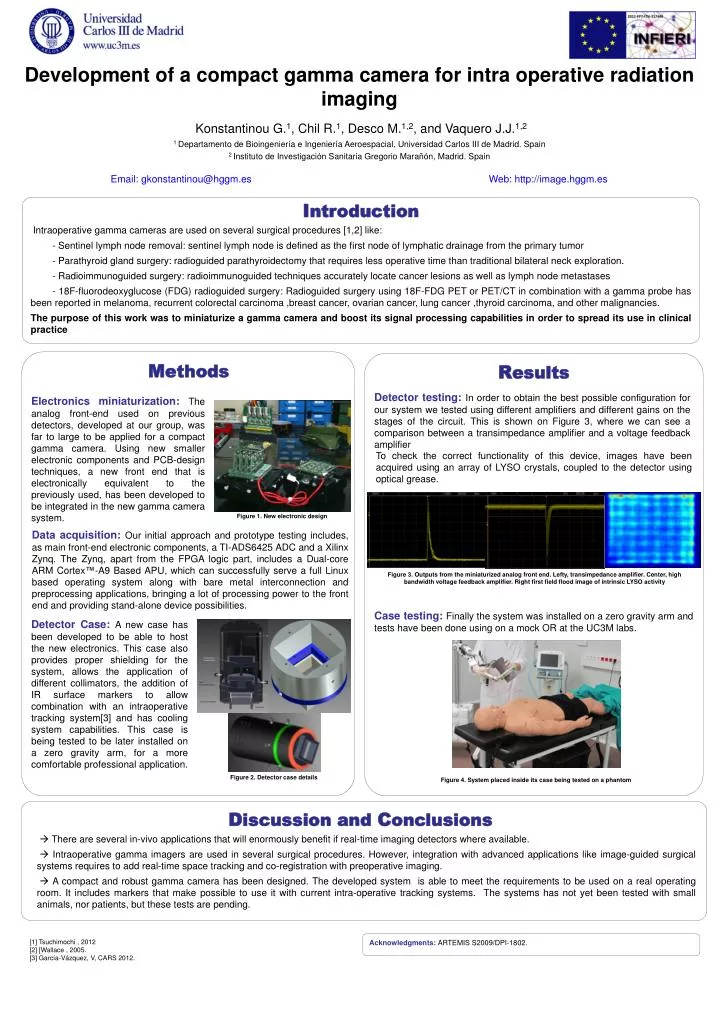
Development of a compact gamma camera for intra operative radiation imaging Konstantinou G.1, Chil R.1, Desco M.1,2, and Vaquero J.J.1,2 1 Departamento de Bioingeniería e Ingeniería Aeroespacial, Universidad Carlos III de Madrid. Spain 2 Instituto de Investigación Sanitaria Gregorio Marañón, Madrid. Spain Email: gkonstantinou@hggm.es Web: http://image.hggm.es Introduction Intraoperative gamma cameras are used on several surgical procedures [1,2] like: - Sentinel lymph node removal: sentinel lymph node is defined as the first node of lymphatic drainage from the primary tumor - Parathyroid gland surgery: radioguided parathyroidectomy that requires less operative time than traditional bilateral neck exploration. - Radioimmunoguided surgery: radioimmunoguided techniques accurately locate cancerlesions as well as lymph node metastases - 18F-fluorodeoxyglucose (FDG) radioguided surgery: Radioguided surgery using 18F-FDG PET or PET/CT in combination with a gamma probe has been reported in melanoma, recurrent colorectal carcinoma ,breast cancer, ovarian cancer, lung cancer ,thyroid carcinoma, and other malignancies. The purpose of this work was to miniaturize a gamma camera and boost its signal processing capabilities in order to spread its use in clinical practice Methods Results Electronics miniaturization: The analog front-end used on previous detectors, developed at our group, was far to large to be applied for a compact gamma camera. Using new smaller electronic components and PCB-design techniques, a new front end that is electronically equivalent to the previously used, has been developed to be integrated in the new gamma camera system. Detector testing: In order to obtain the best possible configuration for our system we tested using different amplifiers and different gains on the stages of the circuit. This is shown on Figure 3, where we can see a comparison between a transimpedance amplifier and a voltage feedback amplifier To check the correct functionality of this device, images have been acquired using an array of LYSO crystals, coupled to the detector using optical grease. Figure 1. New electronic design Data acquisition: Our initial approach and prototype testing includes, as main front-end electronic components, a TI-ADS6425 ADC and a Xilinx Zynq. The Zynq, apart from the FPGA logic part, includes a Dual-core ARM Cortex™-A9 Based APU, which can successfully serve a full Linux based operating system along with bare metal interconnection and preprocessing applications, bringing a lot of processing power to the front end and providing stand-alone device possibilities. Figure 3. Outputs from the miniaturized analog front end. Lefty, transimpedance amplifier. Center, high bandwidth voltage feedback amplifier. Right first field flood image of intrinsic LYSO activity Case testing: Finally the system was installed on a zero gravity arm and tests have been done using on a mock OR at the UC3M labs. Detector Case: A new case has been developed to be able to host the new electronics. This case also provides proper shielding for the system, allows the application of different collimators, the addition of IR surface markers to allow combination with an intraoperative tracking system[3] and has cooling system capabilities. This case is being tested to be later installed on a zero gravity arm, for a more comfortable professional application. Figure 2. Detector case details Figure 4. System placed inside its case being tested on a phantom Discussion and Conclusions There are several in-vivo applications that will enormously benefit if real-time imaging detectors where available. Intraoperative gamma imagers are used in several surgical procedures. However, integration with advanced applications like image-guided surgical systems requires to add real-time space tracking and co-registration with preoperative imaging. A compact and robust gamma camera has been designed. The developed system is able to meet the requirements to be used on a real operating room. It includes markers that make possible to use it with current intra-operative tracking systems. The systems has not yet been tested with small animals, nor patients, but these tests are pending. [1] Tsuchimochi, 2012 [2] [Wallace , 2005. [3] García-Vázquez, V, CARS 2012. Acknowledgments: ARTEMIS S2009/DPI-1802.