Download
1 / 5
60 likes | 353 Views
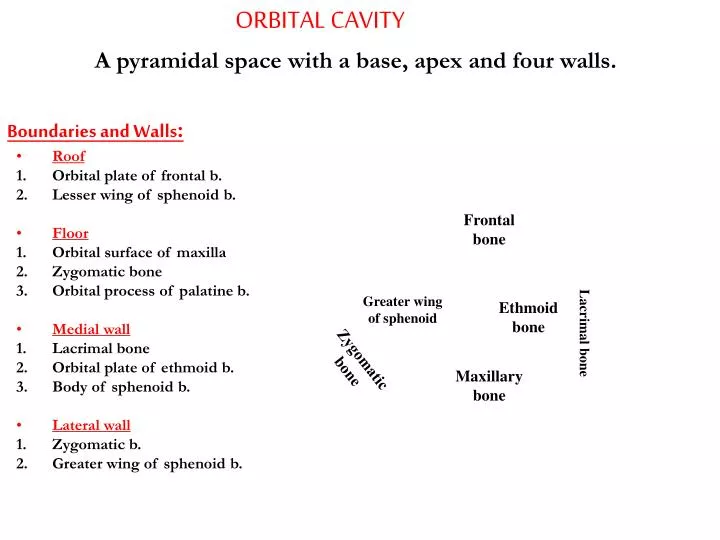
ORBITAL CAVITY. A pyramidal space with a base, apex and four walls. Boundaries and Walls : Roof Orbital plate of frontal b. Lesser wing of sphenoid b. Floor Orbital surface of maxilla Zygomatic bone Orbital process of palatine b. Medial wall Lacrimal bone Orbital plate of ethmoid b.

E N D
ORBITAL CAVITY A pyramidal space with a base, apex and four walls. • Boundaries and Walls: • Roof • Orbital plate of frontal b. • Lesser wing of sphenoid b. • Floor • Orbital surface of maxilla • Zygomatic bone • Orbital process of palatine b. • Medial wall • Lacrimal bone • Orbital plate of ethmoid b. • Body of sphenoid b. • Lateral wall • Zygomatic b. • Greater wing of sphenoid b. Frontal bone Greater wing of sphenoid Ethmoid bone Lacrimal bone Zygomatic bone Maxillary bone
Some Features of The Orbit • Lacrimal fossa (1) (lodges the orbital part of the lacrimal gland) • Lacrimal groove and crests (2) (lodges the lacrimal sac) • Trochlear fovea (3) (attachment of the trochlea) • Ant. and post. ethmoidal canals (4) (ant. and post. ethmoidal nerves and vessels) • Zygomatico-facial and Z-temporal canals (zygomaticofacial and z-temporal n. and v.) • Optic canal (5) (optic nerve, ophthalmic a. & meningeal sheath) • Supraorbital foramen (6) (supraorbital n. and v.) • Infraorbital groove, canal and foramen (7) (infraorbital n. and v.) • Superior orbital fissure (8) • Inferior orbital fissure (9) 6 3 1 4 5 8 2 9 7 7
Communications of the Orbit • The orbital cavity communicates with the: • Anterior cranial fossa viaant. and post. ethmoidal canals (1) • Middle cranial fossa viaoptic canal and the superior orbital fissure (2, 3) • Infratemporal fossa viainferior orbital fissure (4) • Nasal cavity vianasolacrimal canal (5) 1 2 3 5 4
Contents of the Orbital Cavity • Orbital fat • Eyeball • Muscles • Vessels • Nerves • Ciliary ganglion • Lacrimal gland Muscles of the Orbit Levator palpebrae suprioris Extrinsic muscles of the eye Intrinsic muscles of the eye • 4 Recti muscles • Superior rectus • Inferior rectus • Medial rectus • Lateral rectus • Dilator pupillae • Constrictor pupillae • Ciliary muscle • 2 Oblique muscle • Superior oblique • Inferior oblique
Levator palpebrae superioris m. O: roof of the orbit in front of the optic canal I : superficial lamella: front of superior tarsus & skin of upper eyelid deep lamella: upper border of superior tarsus & superior fornix of conjunctiva A: elevation of upper eyelid and sup fornix of conjunctiva N: sup division of oculomotor n. Levator palp sup Lev Palp Sup Insertion of lev palp sup Superior tarsus