Download
1 / 61
610 likes | 792 Views
Heart & Circulation. Circulation. The human circulatory system consists of 96,000 km of blood vessels that transport blood to each cell in the body. Your entire blood volume (about 5L) is pumped every minute. . The circulatory system performs the following functions:.

E N D
Circulation The human circulatory system consists of 96,000 km of blood vessels that transport blood to each cell in the body. Your entire blood volume (about 5L) is pumped every minute.
The circulatory system performs the following functions: 1. carries oxygen and nutrients to the cells 2. carries carbon dioxide and waste away from the cells 3. carries hormones to target organs 4. distributes heat throughout the body 5. helps defense of invading micro-organisms
Blood Vessels • Arteries – carry blood away from the heart. Their thick walls are composed of three distinct, elastic layers. Each time the heart pumps, the arteries stretch to accommodate the rush of blood. This is felt in the neck, or on the wrist as a pulse. • Arterioles - are smaller arteries whose middle layer is composed of elastic fibers and smooth muscle. The arterioles are able to contract and relax, controlling blood flow to different parts of the body.
Vasoconstriction – the narrowing of blood vessels, decreasing flow to the tissues.
Capillaries – are tiny blood vessels composed of a single layer of cells. This is the site of fluid and gas exchange between the cells and the body tissues. Many capillaries are only as thick in diameter as one red blood cell (<0.005 mm). Pressure in the capillaries is high, increasing the risk of rupturing and causing a bruise.
Venules – larger blood vessels that form as capillaries merge. The venules are lined with smooth muscle to ensure blood continues to flow back towards the heart. • Veins – larger blood vessels that result as venules merge, take blood back towards the heart. Veins also serve as blood reservoirs, holding up to 65% of the total blood volume. Blood pressure in the veins is quite low, so the veins have uni-directional valves that ensure the one way flow of blood. Skeletal muscles also help aid venous flow. Venous pressure increases when skeletal muscles contract and push on the vein, forcing blood upwards.
Problems with blood vessels: • Aneurysm – a bulge or weakening in the wall of a blood vessel. • Atherosclerosis – degeneration of blood vessels caused by the accumulation of fat deposits (plaque) in the inner wall.
Bruising – rupture of capillary beds cause blood to leak into the extra-cellular space. • Varicose Veins – damage to the one-way valves in veins causes blood to pool and the veins to bulge.
Circulation • The heart consists of two parallel pumps. The right connects to blood vessels that circulate blood to the lungs, for oxygenation, and back to the heart. This system is called the pulmonary circulatory system. The second, left hand pump, connects blood vessels to the body and circulates blood to the body tissues. This system is called the systemic circulatory system. One way blood flow is maintained by uni-directional valves in the heart and in the blood vessels.
Pulmonary System Systemic System
Closed Circulation • Blood never leaves vessels
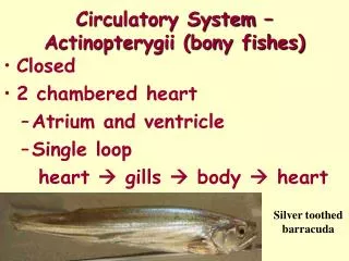
Double Circulatory System • In mammals, birds and reptiles, the blood is pumped twice before returning to its origin • Fish, and other lower organisms pump the blood only once
Four Chambered Heart • oxygenated and deoxygenated blood do not mix in the heart • septum separates the right and left sides
Heart Chambers • The top chambers are called atria (sing. Atrium) • These collect blood from major veins and pump it into the bottom chambers
Ventricles • Much larger and more muscular than atria • Pump blood into arteries for distribution to body (and lungs)
Vessels • Arteries – carry blood away from heart • Veins – carry blood toward heart • Systemic circulation – blood flow to and from body organs (not lungs) • Pulmonary circulation – blood flow to and from lungs
Atrioventricular Valves • Separate the atria and ventricles
Atrioventricular valves have: • Chordae tendinae prevent flaps from everting (opening backward) • Papillary muscles to attach chordae tendinae to ventricle wall
The Bicuspid Valve (AV) • Also called the mitral valve • On the left side of the heart • Has two flaps
Tricuspid Valve (AV) • On the right side • Has three flaps
Semilunar Valves • At the entrance to the major arteries are smaller valves with no muscular attachments • These have three flaps each and prevent backflow into the ventricles
Valve movie • http://www.wellesley.edu/Biology/Courses/111/HeartValves.MOV • video
Pericardium • Membrane around heart which prevents friction between heart and lungs • Also helps isolate infection
Coronary Arteries • The aorta branches and one of the branches comes back to serve the heart muscle (myocardium)
Coronary Circulation • These arteries can become blocked with plaque (cholesterol and calcium deposits) and clots can occur causing: • pain: angina pectoris • Heart muscle death: myocardial infarction (heart attack)
Risks for Heart Disease • Genetics • Male • Smoking • Obesity • Diet (saturated fat and cholesterol) • Lack of exercise
Heart Beats • Cardiac Muscle: striated, branched • Is myogenic muscle: can contract without nerve impulse
Heart Beats • Chemoreceptors in aorta and carotid (neck) arteries detect high carbon dioxide levels in blood (lesser extent – oxygen is also monitored) • Cranial nerves carry this information to the heart
Heart Beats The heart beat is controlled by the sympathetic (stimulating) and parasympathetic (relaxing) branches of the nervous system.
Heart Beats Tempo is set by the sinoatrial node (SA node) : the pacemaker
Heart Beats • Electrochemical impulses from the S.A. node cause contraction of atria muscle
Heart Beats • the impulse travels to another cluster of nervous tissue – the Atrioventricular node (A.V. node)
Heart Beats • The A.V. node sends impulses through Purkinje fibers to all parts of the ventricles • Ventricles contract simultaneously
Heart Beats • Heart muscle then ‘rests’ before the cycle begins again • ‘systole’ – phase of heart contraction • ‘diastole’ – phase of heart relaxation • ‘lub – dub’ – heart sounds caused by valves slamming shut
Heart Beats • Normal Heart Rate – 80 beats/minute • Bradycardia - < 50 beats/min • Tachycardia - > 100 beats/min
Monitoring Heart Beat • EKG (or ECG) – electrocardiogram • Uses electrical activity in heart muscle to diagnose problems
EKG • P wave – depolarization preceding atrial contraction • QRS complex – precedes ventricular • T wave – repolarization of myocardium
Defibrillator • Used to shock hearts into proper rhythm
Other Abnormal Rhythms • tachycardia