Download
1 / 1
10 likes | 111 Views
Introduction. a. b. a. b. d. c. Methods. Acknowledgements. Identification of the Mechanism for Chiral Modification of Calcite Morphology by Aspartic Acid Enantiomers Rachel L. Reese and Sausan Jaber (faculty advisor: Dr. Ryan E. Sours)

E N D
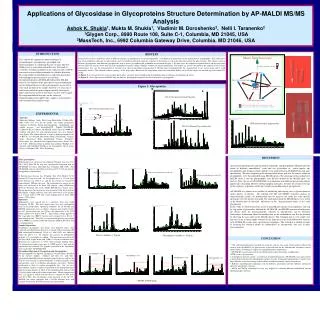
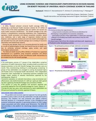
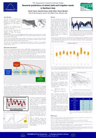
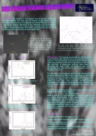
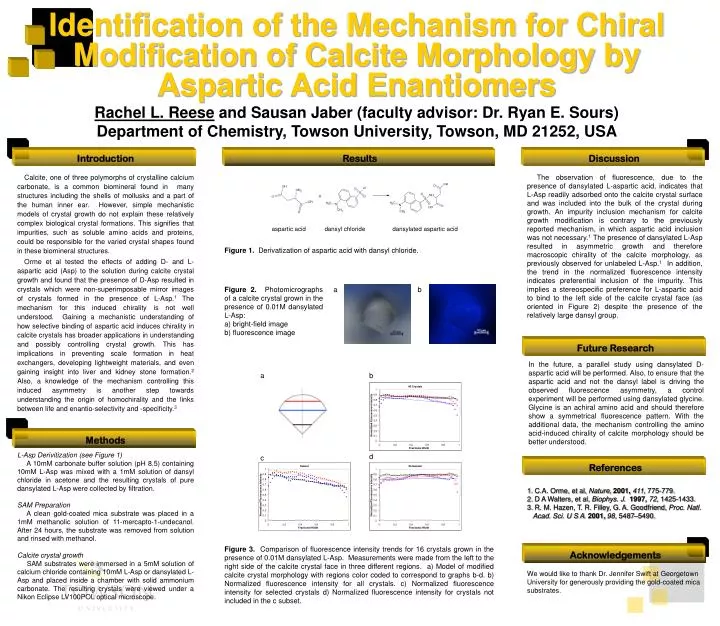
Introduction a b a b d c Methods Acknowledgements Identification of the Mechanism for Chiral Modification of Calcite Morphology by Aspartic Acid Enantiomers Rachel L. Reese and Sausan Jaber (faculty advisor: Dr. Ryan E. Sours) Department of Chemistry, Towson University, Towson, MD 21252, USA Results Discussion Calcite, one of three polymorphs of crystalline calcium carbonate, is a common biomineral found in many structures including the shells of mollusks and a part of the human inner ear. However, simple mechanistic models of crystal growth do not explain these relatively complex biological crystal formations. This signifies that impurities, such as soluble amino acids and proteins, could be responsible for the varied crystal shapes found in these biomineral structures. Orme et al tested the effects of adding D- and L- aspartic acid (Asp) to the solution during calcite crystal growth and found that the presence of D-Asp resulted in crystals which were non-superimposable mirror images of crystals formed in the presence of L-Asp.1 The mechanism for this induced chirality is not well understood. Gaining a mechanistic understanding of how selective binding of aspartic acid induces chirality in calcite crystals has broader applications in understanding and possibly controlling crystal growth. This has implications in preventing scale formation in heat exchangers, developing lightweight materials, and even gaining insight into liver and kidney stone formation.2 Also, a knowledge of the mechanism controlling this induced asymmetry is another step towards understanding the origin of homochirality and the links between life and enantio-selectivity and -specificity.3 The observation of fluorescence, due to the presence of dansylated L-aspartic acid, indicates that L-Asp readily adsorbed onto the calcite crystal surface and was included into the bulk of the crystal during growth. An impurity inclusion mechanism for calcite growth modification is contrary to the previously reported mechanism, in which aspartic acid inclusion was not necessary.1 The presence of dansylated L-Asp resulted in asymmetric growth and therefore macroscopic chirality of the calcite morphology, as previously observed for unlabeled L-Asp.1 In addition, the trend in the normalized fluorescence intensity indicates preferential inclusion of the impurity. This implies a stereospecific preference for L-aspartic acid to bind to the left side of the calcite crystal face (as oriented in Figure 2) despite the presence of the relatively large dansyl group. aspartic acid dansyl chloride dansylated aspartic acid Figure 1. Derivatization of aspartic acid with dansyl chloride. • Figure 2. Photomicrographs of a calcite crystal grown in the presence of 0.01M dansylated L-Asp: • bright-field image • fluorescence image Future Research In the future, a parallel study using dansylated D-aspartic acid will be performed. Also, to ensure that the aspartic acid and not the dansyl label is driving the observed fluorescence asymmetry, a control experiment will be performed using dansylated glycine. Glycine is an achiral amino acid and should therefore show a symmetrical fluorescence pattern. With the additional data, the mechanism controlling the amino acid-induced chirality of calcite morphology should be better understood. L-Asp Derivitization (see Figure 1) A 10mM carbonate buffer solution (pH 8.5) containing 10mM L-Asp was mixed with a 1mM solution of dansyl chloride in acetone and the resulting crystals of pure dansylated L-Asp were collected by filtration. SAM Preparation A clean gold-coated mica substrate was placed in a 1mM methanolic solution of 11-mercapto-1-undecanol. After 24 hours, the substrate was removed from solution and rinsed with methanol. Calcite crystal growth SAM substrates were immersed in a 5mM solution of calcium chloride containing 10mM L-Asp or dansylated L-Asp and placed inside a chamber with solid ammonium carbonate. The resulting crystals were viewed under a Nikon Eclipse LV100POL optical microscope. References • C.A. Orme, et al, Nature, 2001,411, 775-779. • D A Walters, et al, Biophys. J. 1997,72, 1425-1433. • R. M. Hazen, T. R. Filley, G. A. Goodfriend, Proc. Natl. Acad. Sci. U S A. 2001,98, 5487–5490. Figure 3. Comparison of fluorescence intensity trends for 16 crystals grown in the presence of 0.01M dansylated L-Asp. Measurements were made from the left to the right side of the calcite crystal face in three different regions. a) Model of modified calcite crystal morphology with regions color coded to correspond to graphs b-d. b) Normalized fluorescence intensity for all crystals. c) Normalized fluorescence intensity for selected crystals d) Normalized fluorescence intensity for crystals not included in the c subset. We would like to thank Dr. Jennifer Swift at Georgetown University for generously providing the gold-coated mica substrates.