Download
1 / 78
790 likes | 803 Views
The Pathology of Intestines I. Developmental anomalies. Atresia (bowel): complete failure of development of the intestinal lumen ( imperforate anus ) Stenosis (bowel): narrowing of the intestinal lumen.

E N D
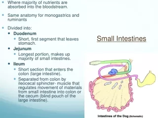
Developmental anomalies • Atresia(bowel): complete failure of development of the intestinal lumen (imperforate anus) • Stenosis(bowel): narrowing of the intestinal lumen
Duplication (small intestine): well-formed saccular-tubular cystic spaces (may or may not communicate with the lumen). • Omphalocele(small intestine): a membranous sac; herniation. • Malrotation (bowel): malposition of the large intestinal components (caecum in the left upper quadrant).
Meckel diverticulum: (small intestine) • Common; in ileum; • Failure of involution of the omphalomesenteric (vitelline) duct which connects the lumen of the developing gut to the yolk sac; • Persistent blind-ended tubular protrusion (5-6 cm long); • Contains all three layers of the normal bowel wall: mucosa, submucosa, and muscularis propria; • Asymptomatic • Pernicious anemia-like syndromes (bacteria B12 depletion); • Acute appendicitis-like syndrome (heterotopic rests of gastric mucosa Peptic ulcerationbleeding).
Hirschsprung’s Disease(Congenital Megacolon) Pathogenesis: The enteric neuronal plexus develops from neural crest cells which must migrate into the bowel wall during developmentmostly in a cephalad-to-caudad direction. Congenital megacolon, or Hirschsprung’s disease, results when the migration of neural crest cells arrests at some point before reaching the anus. Hence a segment remains that lacks both Meissner’s submucosal and Auerbach’s myenteric plexuses. Loss of enteric neuronal coordination leads to (1) functional obstruction (2) colonic dilatation proximal to the affected segment. Occurs in approximately 1 out of 5000 to 8000 live births M/F : 4/1
Hirschsprung’s disease is characterized by the absence of • ganglion cells, • ganglia, in the muscle wall and submucosa of the affected segment. • The rectum is always affected, (most cases involve the rectum and sigmoid only), • Proximal to the aganglionic segment, the colon undergoes progressive dilatation and hypertrophy. • With time, the colon may become massively distended, sometimes achieving a diameter of 15 to 20 cm (megacolon) • The colonic wall becomes markedly thinned and may rupture, usually near the caecum. • Mucosal inflammation or shallow stercoral ulcers produced by impactedfeces may appear. • Enterocolitis.
(1) Chagas’ disease (in which the trypanosomes directly invade the bowel wall to destroy the enteric plexuses); (2) obstruction of the bowel as by a neoplasm or inflammatory stricture; (3) toxic megacolon complicating ulcerative colitis or Crohn’s disease (4) a functional psychosomatic disorder. Acquired megacolon
Ischemic Bowel Disease Arterial thrombosis Arterial embolism Venous thrombosis Nonocclusive ischemia Miscellaneous: radiation injury, volvulus, stricture, and internal or external herniation. Angiodysplasia Hemorrhoids Vascular disorders
Ischemic Bowel Disease • Restricted to the small or large intestine or may affect both, • Acute occlusion of one of the three major supply trunks of the intestines(celiac and superior and inferior mesenteric arteries)infarction. • Lesions within the end-arteries, which penetrate the gut wall, produce small, focal ischemic lesions.
The severity of injury ranges from • (1)transmural infarction of the gut, involving all visceral layers (acute occlusion of a major artery); • (2)mural infarction of the mucosa and submucosa; • (3)mucosal infarction (erosions), if the lesion extends no deeper than the muscularis mucosa.
Arterial embolism: • cardiac vegetations, • angiographic procedures, • aortic thromboembolism. • Arterial thrombosis: • severe atherosclerosis (a.mesenterica), • systemic vasculitis (polyarteritis nodosa), • dissecting aneurysm, • angiographic procedures, • aortic reconstructive surgery, • surgical accidents, • hypercoagulable states, • oral contraceptives.
Venous thrombosis: • hypercoagulable states, • oral contraceptives, • antithrombin III deficiency, • intraperitoneal sepsis, • the postoperative state, • invasive neoplasms (particularly hepatocellular carcinoma), • cirrhosis, • abdominal trauma. • Nonocclusive ischemia: • cardiac failure, • shock, • dehydration, • vasoconstrictive drugs (digitalis, vasopressin, cocaine, heroin). • Miscellaneous: • Radiation • Volvulus • Stricture • herniation
Morphology Transmural Infarction: • Sudden and total occlusion of mesenteric arterial blood intestinal infarction. • Arterial or venous occlusionhemorrhagic infarct, • ischemic injury mucosal necrosis 18-24 h fibrinous exudate over the serosa • Margins of the infarct: • in arterial occlusions : distinct (demarcation) • in venous occlusions : less distinct • Microscopy: edema, interstitial hemorrhage, necrosis • (24 h)gangreneperforationperitonitis !!!
Mural & Mucosal Infarction: • In any level of the gut from the stomach to anus • lesions may be multifocal-scattered or continuous-widely distributed (depends on the level of the arterial narrowing), • does not affect the entire thickness (may not be visible from the serosal surface), • on opening the bowel, there is hemorrhagic, edematous thickening of the mucosa. • Superficial ulcerations with • edema, • hemorrhage, • fibrinous inflammation (psedomembrane due to superinfection).
Chronic Ischemia: • Chronic vascular insufficiency mucosal inflammation and ulceration • Submucosal chronic inflammation and fibrosis stricture. • Segmental and patchy.
Angiodysplasia • Tortuous dilatations of submucosal and mucosal blood vessels • most often in the cecum or right colon • after the sixth decade of life • intestinal bleeding: • chronic and intermittent (anemia) • acute and massive.
Hemorrhoids • Variceal dilatations of the anal and perianal venous plexuses • Persitently elevated venous pressure within the hemorrhoidal plexus • Predispositions: • chronic constipation, • pregnancy. • Thin-walled, dilated vessels • Complications: Bleeding, prolapsing.
Diarrheal diseases • Diarrhea is an increase in stool: • Mass • Frequency • Fludity • Disentery is a kind of diarrhea with: • Low-volume • Pain • Hemorrhage
Diarrhea • 1. Secretory diarrhea • 2. Osmotic diarrhea • 3. Exudative diarrhea • 4. Malabsorption • 5. Deranged motility
1. Secretory diarrhea • Intestinal fluid secretion (catarrh) • Serous • Causes: • Infection (bacteria, virus) • Neoplastic (secretion of peptides and serotonin) • Excessice laxative use
2. Osmotic diarrhea • Excessive osmotic forces • Causes: • Lactulose therapy (hepatic encephalopathy, constipation) • Gut lavage • Antacids (magnesium salts)
3. Exudative diarrhea • Purulent/bloody stool • Causes: • Infections • escherichia, • campylobacter, • shigella, • salmonella, • Entamoeba histolytica, • Idiopathic inflammatory bowel disease
4. Malabsorption • Voluminous, bulky stool • Causes: • Defective intraluminal digestion • Defective mucosal cell absorption • Reduced small intestinal surface area • Lymphatic obstruction • Infection (Giardia)
5. Deranged motility • Decreased intestinal retention time • Surgical reduction of gut length • Neural dysfunction (irritable bowel syndrome) • Hyperthyroidism • Decreased motility • Surgery • Bacterial overgrowth in the small intestine
Infectious enterocolitis • Intestinal diseases of microbial origin • Diarrhea and sometimes ulceroinflammatory changes • Most common offenders • rotavirus • Norwalk virus • Enterotoxigenic Escherichia coli
Offenders vary with the • age, • nutrition, • immune status of the host, • environment (living conditions, public health measures), • Special predispositions: • hospitalization, • wartime dislocation, • foreign travel.
Viral Gastroenterocolitis • The small intestinal mucosa usually exhibits • shortened villi • infiltration of the lamina propria by lymphocytes • vacuolization and loss of the microvillus brush border in surface epithelial cells • crypts appear hypertrophied • viral particles within surface epithelial cells by electron microscopy and in stool.
Bacterial Gastroenterocolitis • Numerous bacteria and several pathogenic mechanisms: • Ingestion of preformed toxin, present in contaminated food (major offenders are Staphylococcus aureus, Vibrios, and Clostridium perfringens) • Infection by toxigenic organisms (which proliferate within the gut lumen and elaborate an enterotoxin) • Infection by enteroinvasive organisms (which proliferate, invade, and destroy mucosal epithelial cells)
Most bacterial infections exhibit a general nonspecific pattern: • damage of the surface epithelium • decreased epithelial cell maturation • an increased mitotic rate (“regenerative change”) • hyperemia and edema of the lamina propria • variable neutrophilic infiltration into the lamina propria and epithelial layer.
Salmonella (multiple species, including S. typhimurium and S. paratyphi): • primarily ileum and colon • blunted villi, • vascular congestion, • Peyer’s patch involvement with swelling, • congestion, • ulceration (linear ulcers) • Typhoid fever : may result in chronic infection of • biliary tree, • joints, • bones, • meninges.
Shigella • primarily distal colon • acute mucosal inflammation and erosion • purulent exudate • Campylobacter • small intestine, appendix, colon • villus blunting • multiple superficial ulcers • mucosal inflammation • purulent exudate
Yersinia enterocolitica and Y. pseudotuberculosis: • ileum, appendix, and colon • mucosal hemorrhage and ulceration • bowel wall thickening • Peyer’s patch and mesenteric lymph node hypertrophy with necrotizing granulomas • systemic spread (with peritonitis, pharyngitis, pericarditis 3-Ps) • V. cholerae: • essentially intact small intestinal mucosa, • with mucus-depleted crypts • C. perfringens: • similar to V. cholerae but with some epithelial damage; • some strains produce a severe necrotizing enterocolitis with perforation.
Necrotizing Enterocolitis Neonates (premature or of low birth weight) • acute, necrotizing inflammation. • A combination of • ischemic injury, • colonization by pathogenic organisms, • excess protein substrate in the intestinal lumen, • functional immaturity of the neonatal gut.
The disease may present as a mild gastrointestinal disturbance or as a fulminant illness with • intestinal gangrene, • perforation, • sepsis, • shock. • Terminal ileum and ascending colon, • although in severe cases, the entire small and large bowel may be involved.
In early phases, the mucosa exhibits • edema, • hemorrhage, • necrosis • As the disease progresses, the full thickness of the bowel wall becomes • hemorrhagic, • inflamed, • gangrenous • frank intraluminal hemorrhage • mural gas formation • Reparative changes • epithelial regeneration • granulation tissue formation • fibrosis.
Antibiotic-Associated Colitis (Pseudomembranous Colitis) • C. difficile (a normal gut commensal) • acute colitis • plaque-like adhesion of fibrinopurulent-necrotic debris and mucus to damaged colonic mucosa
following a course of broad-spectrum antibiotic therapy • also may occur following any severe mucosal injury, • ischemic colitis, • volvulus, • necrotizing infections (staphylococci, shigella, candida, necrotizing enterocolitis)
Malabsorption Syndromes • Malabsorption is characterized by suboptimal absorption of fats, fat-soluble and other vitamins, proteins, carbohydrates, electrolytes and minerals, and water. • The consequences of malabsorption affect many organ systems.
Etiology Mal-digestion: Exocrine pancreatic disease Lack of bile salts Disaccharidase (lactase, etc.) deficiency Problems with the small bowel mucosa: Sprue Crohn's disease Whipple's disease Acute infections Parasites (Giardia) Allergic gastroenteritis Amyloidosis Lymphomas Radiation sickness / B12 / folate deficiency Super-fast transit time: Laxatives Cholera Vasoactive intestinal polypeptide-producing tumors Mechanical problems: Blocked lymphatics (cancer, TB) After re-routing surgery (gastrectomy, bypass)
Clinicopathology Alimentary tract: diarrhea abdominal pain weight loss vitamin deficiencies Hematopoietic system: anemia (iron, pyridoxine, folate, or vitamin B12 deficiency) bleeding (vitamin K deficiency) Nervous system: peripheral neuropathy Skin: purpura and petechiae edema dermatitis hyperkeratosis Musculoskeletal system: osteoporosis tetany Endocrine system: amenorrhea impotence infertility hyperparathyroidism
Celiac sprue • Small intestine • idiosyncratic reaction to gliadin, a protein in the gluten of wheat, rye, and barley • an antibody against the transglutaminase • activated cytotoxic killer-T cells invade the epithelium • Microscopy: • villi disappear • crypts deepen
Whipple'sdisease • Small intestine & systemic • lipid pools in the mucosa • Tropheryma whippelii(~actinomyces) • bacilli-laden macrophages in: • gut mucosa • lymph nodes, • joints, • endocardium • brain.


![STOMACH, INTESTINES, RECTUM [SURGICOSE]](https://cdn4.slideserve.com/8061806/medical-instruments-medical-instruments-dt.jpg)