Download
1 / 10
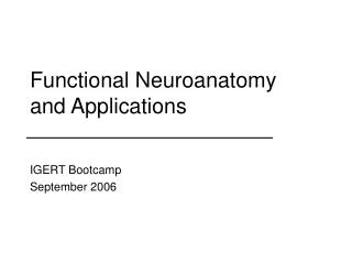
130 likes | 684 Views
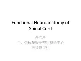
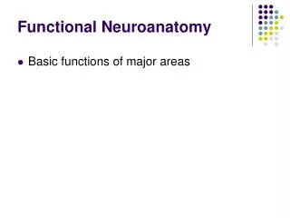
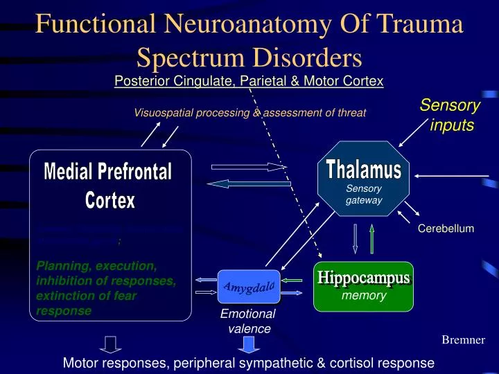
Thalamus. Sensory gateway. Functional Neuroanatomy Of Trauma Spectrum Disorders. Posterior Cingulate, Parietal & Motor Cortex. Sensory inputs. Visuospatial processing & assessment of threat. Medial Prefrontal Cortex. Cerebellum. Anterior Cingulate, orbitofrontal, subcallosal gyrus ;

E N D
Thalamus Sensory gateway Functional Neuroanatomy Of Trauma Spectrum Disorders Posterior Cingulate, Parietal & Motor Cortex Sensory inputs Visuospatial processing & assessment of threat Medial Prefrontal Cortex Cerebellum Anterior Cingulate, orbitofrontal, subcallosal gyrus; Planning, execution, inhibition of responses, extinction of fearresponse Hippocampus Amygdala memory Emotional valence Bremner Motor responses, peripheral sympathetic & cortisol response
Spaced exposure causes incubation acutely Sequential Day 2 Cue Exposures Mark Barad, et al. (2000).
Biological Studies of Child and Adolescent Traumatic Stress • Structural brain development • Neurophysiology • Neurohormones
CRH – peptide corticotropin hormone Mediates autonomic, immune, behavioral components of stress responses Important role in anxiety and fear Intraventricular administration of CRH – behavioral changes * Suppression of exploratory behavior * Potentiation of the acoustic startle reflex * Facilitation of fear conditioning (Meaney, et al.)
CORTISOL RESPONSE Abused girls have higher a.m. cortisol, lower in p.m., more overall cortisol secretion PUTNAM’S PROSPECTIVE STUDY
Hippocampal Volume Reduction In PTSD NORMAL PTSD MRI scan of the hippocampus in a normal control & patient with PTSD secondary to childhood abuse. The hippocampus, outlined in red, is visibly smaller in PTSD. Overall 12% reduction in volume in PTSD. Bremner et al. Am J Psychiatry. 1995; 152:973-981. Bremner et al. Biol Psychiatry. 1997; 41:23-32.
Normal Developmental Course of Startle Inhibition • Prior to about 4 years of age, inhibitory startle modulation is weak or absent, whereas facilitatory startle modulation to sustained prestimulation is strong in early infancy. • Both inhibitory and facilitatory startle modulation show, respectively, peak losses in startle inhibition and peak gains in startle facilitation at about 4.5 years of age. • Progressive increase in inhibition and decrease in facilitation follow until, by 8 years of age, mature values are obtained.
Modulation of Startle Amplitude by Prestimulation in Six Children with PTSD and in Six Control Children 25 – msec Tones Sustained Tones Difference in EMG Amplitudes Between Warned and Unwarned Conditions inhibtion facilitation WARNING INTERVAL (msec) Pynoos, et al., 1997