Download
1 / 26
260 likes | 267 Views
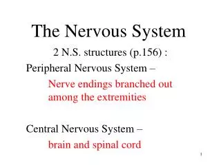
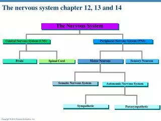
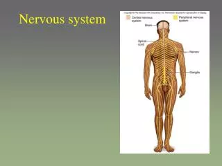
NERVOUS SYSTEM The nervous system can be divided into: A: Central nervous system (CNS) Which includes brain and spinal cord B: Peripheral nervous system (PNS) Which composed of nerve fibers and nerve ganglia and nerve endings C: Autonomic nervous system (ANS)

E N D
NERVOUS SYSTEM The nervous system can be divided into: A: Central nervous system (CNS) Which includes brain and spinal cord B: Peripheral nervous system (PNS) Which composed of nerve fibers and nerve ganglia and nerve endings C: Autonomic nervous system (ANS) Is composed of two part differ anatomically and functionally The sympathetic system The parasympathetic system Central nervous system (CNS) The general structure of (CNS) composed form White and gray matter Meninges (CNS) consists from two pats:
1. Spinal cord Is a slender never column that passes down wards from the brain into the vertebral canal. It consists of 31 segments each of which gives rise to a pair of spinal nerves. In cross-section The spinal cord of oval in shape, poserioly the spinal cord is divided partially into right and left halves by dorsal or posterior medium septum. While anteriorly is a deep longitudinal cleft called anterior ventral medium fissure. The gray matter of spinal cord occupies a central area that is roughly (H) shaped, the upper arms are called posteror horn of gray matter and the lower arms are called anterior horn.Anterior horn. In addition extending throughout the thoracic and lumber segments there is a lateral horn of gray matter. In the horizontal bar of this H-shaped is an opening the central canal which is a remnant of the lumen of the embryonic neural tube, it is lined by ependymal cells. Gray matter contains mainly cell bodies of neurons (multipolat) and unmyelinated fibers. The white matter is located in the periphery of spinal cord and contains myelinated nerve fiber and unmyelinated nerve fiber and oloigodendrocytes. It does not contain neuronal cell bodies.
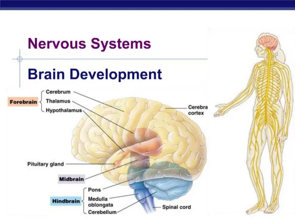
2. Brain In composed of about one hundred billion neurons and nerve fibers, by which the neurons communicate with one another and with neurons of other part of the system. The brain can be divided into; Cerebrum Cerebellum Which is enclosed by skull Cerebrum It is the largest part contains nerve centers associated with sensory and another functions. The cerebrum consists of 2 large masses called (cerebral hemispheres), these spheres are connected by a deep bridge of nerve fibers and are separated by a layer of dura mater. The surface of the cerebrum is marked by numerous ridges is called gray which are separated by grooves which is called sucli. A thin layer of gray matter at the periphery of the cerebral hemispheres is called the cerebral cortex, containing nerve cell, fibers neuralgia and blood vessels. The cerebral cortex is divided into six layers, these are from superficial to deep:
1. Molecular or plexiform layer Composed mainly of nerve fibers, they found between these fibers and the basic part of this layer canals cells which is small nerve cells and possess fusiform shape or stellate shape. 2. Outer granular layer The contains of small triangular nerve cell bodies. 3. Outer pyramidal layer Composed of pyramidal cells and many small granular cells. 4. Inner granular layer It consists of small stellate granule cells. 5. Inner pyramidal layer It contains of large pyramidal cells 6. Multiformn or polymorphic layer It is composed of cells of varying shaped. The most cells in this layer is fusiform cells. The white matter underlying the gray cortex is composed of bundles of myeinated fibers passing in all directions. The cortex of cerebrum responsible for learning memory, initiation of motor response and integration of sensory signals.
Cerebellum Consists of right and left hemispheres and middle lobe called vermis cerebelli. The cerebellum like the cerebrum consists of thin layer of cerebellum cortex on its surface. An thick layer of white matter just undernearth the cortex. The cerebellar cortex on section shows 3 layers: 1. Outer molecular layer A few small nerve cells and many non myelinated fibers tow types of cells found in this layer the stellate and basket cells. 2. central layer of purkinje cells contains the large flask shape which several main dendrites that enter the molecular layer as a fan shaped network which are present only in cerebellum. These cells are only cells that send information to the outside. 3. Inner granular layer Consists of small granule cells The cerebellar cortex is responsible for balance and equilibrium and movement of skeletal muscles. The white matter compose of axon of purkinje cells and climbing fibers and mossy fibers.
In mammals, the main mass of the nerve cells are located in CNS and the cell bodies of neurons usually form groups which are designated. Nuclei, is thy are present in clusters. Laminae, if they are arranged in layers. Columns, if they occur in longitudinal arrange.
The brain and spinal cord are protected by membranes called meninges that are located between the bone and the sift tissue of nervous and they also called cerebrospinal membranes which is consists of connective tissue and it has 3 layers: 1. Dura mater The dura mater is the external layer and is composed of dens connective continuous with the perisoteum of the skull, the dura later that envelops the spinal cord is separated from the periostum of the vertebrae by the epidural space which contains thin walled veins, loss connective tissue and adipose tissue. The dura mater is always separated form the arachnoid by the thin subdurl space the internal surface in the spinal cord is covered by simple quamous epithelial of mesenchymal origin.
2. Arachnoid The arachnoid has two components: A layer in contact with the dura mater. A system of trabecular connecting the layer with the pia mater. The cavities between the trabeculae form the subarachnoid space which is filled with cerebrospinal fluid and is completely from the subdural space. The arachnoid is composed of connective tissue its surfaces are covered by the same type of simple squenmous epithelium that covers the dura mater. In some areas the arachnoid perforates the dura mater forming protrusions that terminate in venous sinuses in the dura mater. These protrusions which are covered by endothelial cells of the veins are called arachnoid villi their function is to reabsorb cerebrospinal fluid into the blood of the venous sinuses. 3. Pia mate The pia mater is a loose connective tissue containing many blood vessels. Although it is located quite close to the nerve tissue It is not in contact with nerve cells or fiber between the pia mater an the neural elements is a thin layer of neuroglial processes adhering firmly to the pia mater. These mater is covered by squamous cells of mesenchymal origin. Blood vessels penetrate the (CNS) through covered by pia mater.
PERIPHERAL NERVOUS SYSTEM (PNS) Which composed of Nerve fibers Ganglia nerve endings 1. Nerve fibers Nerve fibers consist of axons enveloped by a special sheath derived from cells of ectodermal origin. Groups of nerve fibers constitute the tracts of the brain, spinal cord and peripheral nerves exhibit differences in their enveloping sheath. Most axon in adult nerve tissues are covered by single or multiple folds of a sheath cells. In peripheral nerve fibers, the sheath cell is the schwann cell, and in central nerve fibers it is the oligodendrocyte.
The nerve fibers classification into: Myelinated fibers Unmyelinated fibers a. Myelinated fibers It can be used to mean the axon with all of its covers by white fatty sheath or myelin shath. The myelin sheath derived (from plasmalemma of schwann's cell) myelin sheath consists a higher of lipids and proteins. The myelin sheath shows gaps along is path called the (nodes of Ranvier) these represent the spaces between adjacent schwann cells along the length of the axon, the distance between two nodes is called an internode and consists of one schwann cell, the length of the internode varies between (1 and 2mm). There are no schwann cells in the (CNS) the myelin sheath is formed by the processes of the oligodendrocytes. Myelin sheath (white color) is responsible for the color of white matter of (CNS). External to the schwann cells and closely adhering to them is a very delicate connective tissue membrane these called neuro lemma or sheath of schawnns. The schawanns sheath surrounded the myelin sheath in the myelinated fibers.
B. Unmyelinated fibers In both the CNS and PNS not all axons are sheathed in myelin. In ONS all unmyelinted axons are enveloped within simple clefts of the schwann cells and do not have nodes of Ranvier. In CNS is rich in unmyelinated axons unlike those in the peripheral system there axons are not sheathed. There are no schwann cells in CNS. Unmyelinted fibers are responsible for the color of gray matter of CNS.
Structure of the peripheral nerve Are composed of bundles of nerve fibers held together by connective tissue and include spinal nerves connected to spinal cord and cranial nerves connected with brain/ Surrounding the all nerve a sheath of strong connective tissue termed epineurium which is composed of fiberoblasts and collagenous fibers and few elastic fibers. Within the epineurium, nerve fibers are grouped into bundles or fascicles, and each fascicles is surround by connective sheath called perineurim.. Within the perineurium there are strands of delicate connective tissue extending around and between the nerve fiber this is called endoneurium, this is closely adherent to the neurolemma (sheath of schwann) small peripheral nerves lack an epineurium and have only perineurium and endoneurium the peripheral herves conatins on myelin fibers and unmyelinated fibers.
Classification of nerve fibers Nerve fibers are classify functionally into: 1. Sensory (afferent) fibers Fibers carry the information obtained from the interior of the body and the environment to the central nervous system. 2. Motor (efferent) fibers Fibers carry impulses form the CNS to the effector organs commanded by these centers. Nerves possessing only sensory fiber are called sensory nerves. Those composed only of fibers carrying impulses to the effectors are called motor nerves. Most nerves have both sensory and motor fibers and are called mixed nerves these nerves have both myelinated and unmyelinated axons.
2. Ganglia These are collection of cell bodies of neurons located outside the CNS. Ganglia are the 2 main types. A. Sensory ganglia Sensory ganglia receive afferent impulses that go to the CNS two types of sensory ganglia: 1. Cranial or cerebral ganglia Some are associated with cranial nerves. 2. Spinal ganglia Which are associated with the dorsal root of the spinal nerves. Both the ganglia histologically appear as a collection of psedounipolar cell bodies of the afferent neurons.
Sensory ganglia are surrounded by connective tissue capsule composed of fine colagenous and reticular fibers and blood vessels. The connective tissue of capsule continuous directly into dorsal root epineurium and perineurium. The capsule sends connective tissue trabeculae into the ganglion and forms a richly vasculiaized stroma. The connective tissue stroma and bundles of nerve fiber divided the nerve cell bodies into groups of varying size. Each ganglion cell surrounded by a layer of flattened cells called the (capsule cells or satellite cells) which correspond to schwann cells of nerve fibers. A basement membrane separate the layer of satellilte cells from outer layer of connective tissue fiber of fibroblasts.
B. Autonomic ganglia Appear as swelling along the sympathetic chain and within the walls of organs supplied by the autonomic system. Ganglia cells are multi polar usually with several dendrites and single unmyelinated axon. Unlike the sensory ganglia, stroma doesn't show tendency to group and the cell bodies have large rounded nucleus which is light and often a centrically placed. The cell bodies large ganglia are surrounded by a capsule of satellite cells. The satellite cells are fewer and smaller and irregular in arrangement.
Comparison between the sensory ganglia and autonomic ganglia Sensory ganglia (s.g.) is large than autonomic (A.g.). (s.g.) is bursiform but (A.g.) appears as swelling. cell body is psedounipolar in (s.g.) while it is multipolar in (A.g.). In(s.g.) the connective tissue stroma divide the cell body into groups, in (A.g.) don't show tendency to groups. In (s.g.) more satellite cells but the fewer in (A.g.). s.g. nucleus is centric while the A.g. is a centric nucleus.
3. Nerve endings The nerve fibers ending in organs form special modify structure this is called (nerve ending). They are two types of endings classification according of the function Sensory or receptor endings. Motor or effector endings Addition the ending classification according of the tissue type which it found in it. a. Neurmuscular spindle which occur in the striated muscle fibers. b. Motor end plates Found in the striated muscle fibers. c. Disk of merkal Found in the deep of skin epidermis d. Meissner corpuscles Found in skin dermis e. End bulb of Krause Found in skin dermis.
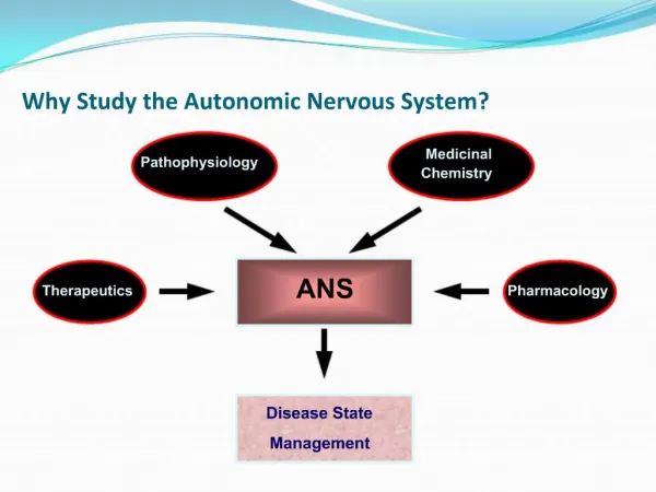
Autonomic nervous system The autonomic nervous system is related to the control of smooth muscle, the secretion of some glands and the modulation of cardiac rhythum. Anatomically it is composed of collections of nerve cells located in the CNS, fibers that leave the CNS through cranial or spinal nerves and nerve ganglia situated in the paths of these fibers. The term autonomic covers al the neural elements concerned with visceral function. In fact the so called autonomic function are as dependent on the central systems as are the motor neurons that trigger muscle contraction. The autonomic nervous system is a two-neuron network. The first neuron of the autonomic chain is located in the CNS. Its axon forms a synape its called preganglionic fibers. And the second neuron multipolar neuron in the chain located in a ganglion of the peripheral outonomic system, it is called postganglionic fibers.
The chemical mediator present in the synaptic vesicles of all pregangloionic ending and at anatomically parasympathetic postganglionic ending is (acetylcholine) which is released from the terminal by nerve impulses. The autonomic nervous system is composed of two parts that differ both anatomically and functional, the sympathetic system and parasympathetic system. 1. Symapathetic system The nuclei (formed by collection of nerve cell bodies) of the sympathetic system are located in the thoracic and lumber segments of spinal cord. Therefore the sympathetic system is also called the thoracolumbar division of the autonomic system. The axons of these neurous-preganglionic fibers leave the CNS by way of the ventral roots. The chemical, mediator of the postganglionic fibers of the sympathetic system is (norepinephrine) which is also produced by the adrenal medulla.
2. Parasympathetic system The parasympathetic system has its nuclei in the medulla and midbrain and in the sacral portion of the spinal cord. The preganglionic fibers of these neurons leave through four of the cranial nerves and also through the second, third an fourth sacral spinal nerves. The parasympathetic system is also called the crainosacral division of the autonomic system. The chemical mediator released by the pre and postganglionic nerve endings of the parasympathetic system.