Download
1 / 33
420 likes | 713 Views
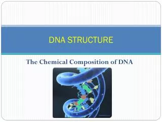
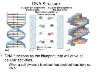
Tertiary Structure: Supercoiled DNA. L = T + W L = Linkage Number number of crossings in planar projection T = Topological winding number- (e.g, number of bp/ 10.5 for B-DNA) W = Writhing number- number of turns the duplex axis makes around itself

E N D
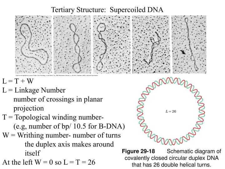
Tertiary Structure: Supercoiled DNA • L = T + W • L = Linkage Number • number of crossings in planar projection • T = Topological winding number- • (e.g, number of bp/ 10.5 for B-DNA) • W = Writhing number- number of turns the duplex axis makes around itself • At the left W = 0 so L = T = 26 Figure 29-18 Schematic diagram of covalently closed circular duplex DNA that has 26 double helical turns.
<95% conversion 5% conversion> of form I to II polyomavirus DNA by DNAse I Proc Natl Acad Sci U S A. 1965 May; 53(5): 1104–1111. J Vinograd, J Lebowitz, R Radloff, R Watson, and P Laipis
Proc Natl Acad Sci U S A. 1965 May; 53(5): 1104–1111. J Vinograd, J Lebowitz, R Radloff, R Watson, and P Laipis
Figure 4.18a: Closed covalent circle Figure 4.18b: Singly-nicked circle Figure 4.20a: Linear double-stranded DNA Tropp: Adapted from Bert, J. M., et al. Biochemistry, Fifth Edition. W.H. Freeman and Company, 2002.
Figure 4.19: Simulated conformation of supercoiled DNA. Tropp:
Figure 4.20b: Relaxed circle Tropp: Adapted from Bert, J. M., et al. Biochemistry, Fifth Edition. W.H. Freeman and Company, 2002.
Figure 4.20d: Unwound circle Tropp: Adapted from Bert, J. M., et al. Biochemistry, Fifth Edition. W.H. Freeman and Company, 2002.
Figure 4.20e: Negative supercoil (right-handed) Tropp: Adapted from Bert, J. M., et al. Biochemistry, Fifth Edition. W.H. Freeman and Company, 2002.
Figure 4.20c: Linear DNA unwound by two right-handed turns Tropp: Adapted from Bert, J. M., et al. Biochemistry, Fifth Edition. W.H. Freeman and Company, 2002.
Figure 4.22b: An underwound covalent circle having only 32 turns of the helix. Figure 4.22a: A nonsupercoiled or relaxed covalent circle having 36 turns of the helix. Figure 4.22c: The molecule in part (b) but with a writhing number of 4 to eliminate the underwinding. Tropp:
Figure 4.21b: Negative superhelix Figure 4.21c: Positive superhelix Tropp: Adapted from Schvartzman, J. B., and Stasiak, A., EMBO Reports 5 (2004): 256-261.
# Steven A. Wasserman and Nicholas R. Cozzarelli # Science, New Series, Vol. 232, No. 4753 (May 23, 1986), pp. 951-960
Figure 4.23: Catalysis of transient breakage of DNA by DNA topoisomerases. Tropp: Adapted from Wang, J. C., Nature Rev. Mol. Cell Biol. 3 (2002): 430-440.
Figure 4.24: Four types of topological coversions catalyzed by topoisomerase I Tropp: Adapted from Kornberg, A., and Baker, T. A. DNA Replication, Second Edition. W.H. Freeman and Company, 1991.
Figure 4.25: Escherichia coli topoisomerase I, a type IA topoisomerase. Tropp: Adapted from Champoux, J. J., Annu. Rev. Biochem. 70 (2001): 369-413.
Figure 4.26: Proposed mechanism of relaxation by E. coli topoisomerase I. Tropp: Adapted from Champoux, J. J., Annu. Rev. Biochem. 70 (2001): 369-413.
Figure 4.26f: Proposed mechanism of relaxation by E. coli topoisomerase I. TroppÑ Adapted from Champoux, J. J., Annu. Rev. Biochem. 70 (2001): 369-413.
Figure 4.27: Proposed mechanism for the catalytic cycle of DNA topoisomerase II. TroppÑ Adapted from Larsen, A. K., et al., Pharmacol. Ther. 2 (2003): 167-181.