Download
1 / 30
310 likes | 499 Views
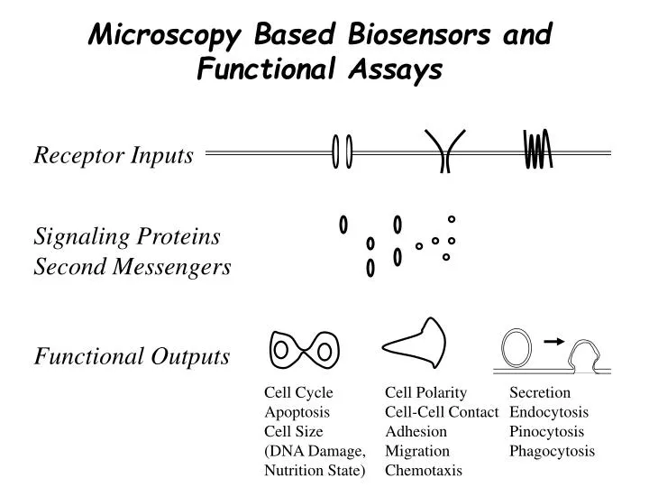
Microscopy Based Biosensors and Functional Assays. Receptor Inputs. Signaling Proteins Second Messengers. Functional Outputs. Cell Cycle Apoptosis Cell Size (DNA Damage, Nutrition State). Cell Polarity Cell-Cell Contact Adhesion Migration Chemotaxis.

E N D
Microscopy Based Biosensors and Functional Assays Receptor Inputs Signaling Proteins Second Messengers Functional Outputs Cell Cycle Apoptosis Cell Size (DNA Damage, Nutrition State) Cell Polarity Cell-Cell Contact Adhesion Migration Chemotaxis Secretion Endocytosis Pinocytosis Phagocytosis
Biosensors, Functional assays Dissecting cellular signaling systems: Perturbations and Biosensors Receptor Inputs Perturbations Functional Outputs
1. Microscopy Strategies to Explore Signaling Systems • Epifluorescence Imaging • Confocal Imaging • Total Internal Reflection Microscopy
A568 YFP YFP YFP YFP YFP YFP YFP Automated assays for receptor endocytosis Anti FLAG M1 monoclonal Ab Internalization (~30%) YFP Epinephrin + EDTA 2mM Fix, perm, 2ndary Ab
Automated immunofluorescence analysis (epifluorescence) CFP-Arf1 (DN) CFP YFP-b2-adrenergic receptor YFP Internalized receptor Texas Red
Reduced endocytosis rates in the presence of some constitutively active small GTPases Ratio I594/IYFP (% CTR) ARF3 ARF1 ARL4 CTR Potentially involved in regulating endocytosis
b2-adrenergic receptor internalization kinetics (confocal imaging) 45 minute movie, YFP-tagged beta2-adrenergic receptor; epinephrine stimulation after 15 minutes
Calcium signals versus PKC translocation (confocal imaging) Calcium- Crimson GFP-PKCg Antigen stimulation of tumor mast cells
Membrane translocation measured by total internal reflection fluorescence microscopy PKC-GFP +Receptor stimulus The evanescent wave field in TIRF imaging
Teflon ring Adherent cells Laser Low magnification projection (9mm x 7mm imaged area) Camera Monitoring plasma membrane translocation in large numbers of cells Evanescent wave Single Cell Array Technology (E-SCAT)
Recording of GFP-C2 domain plasma membrane translocation in many cells PAF ionomycin Plasma membrane 1 Rel. fluorescence Cytosol 0 50 s Teruel and Meyer, Science, 2002
2. Perturbation strategies suitable for microscopy • RNAi • Expression constructs (wt, DN, CA) • Small molecule perturbations (for example induced translocation)
PCR In Vitro Txn In Vitro Dicing and Purification X 24 = 2304 siRNA pools targeted to signaling domain selected proteins Generating d-siRNA sets based on in vitro dicing Large dsRNA r-Dicer * * Pool of dsiRNAs Transfection In vitro Dicer method developed by Jason Myers in Jim Ferrell’s lab Myers, Jones, Meyer and Ferrell Nature Biotech, 2003
CA small GTPases and cell morphology Multiple Local spread RAB22A RAB27A RAB22B RAB27B RABL2B RAB33A RAB11A RAB11B RAB7L1 RAB8B RABRP RAB5C RAB5A RAB6C RAB5B RAB4B RAB1A RAB9L RAB3B RAB3A RAB1B RAB10 RAB13 RAB26 RAB28 SEC4L RAB30 RAB14 RAB25 RAB18 RAB35 RAB21 RAB32 RAB38 RAB23 RAB9 RAB8 RAB6 RAB4 RAB2 RAB7 RAN Lamellipodia Filopodia Eyelashes Rounding Stress fibers Polar Shrunk Multiple kB-RAS1 kB-RAS2 ARFRP1 CDC42h R-RAS3 R-RAS2 RAP1A RAP1B RAP2B RAP2A RHEB2 ARL7B ARL7A ARF4L CDC42 RRP22 R-RAS H-RAS K-RAS N-RAS RHOA RHOG RHOB RHOH RAGA RAGB RHOC RHOD RHOE RALA RALB ARHE SARA RAC3 RHO7 RHO6 AGS1 RAC2 RAC1 SAR1 ARF1 ARF3 ARF5 ARF4 ARF6 ARL5 ARL4 ARL1 ARL3 ARL2 RAD ARHI TC10 GEM REM TCL RIN RIT Heo and Meyer, Cell, 2003
Rac PM translocation induced by a rapamycin analog NIH3T3 cells w. PM-FRB + FKBP-YFP-Rac1(CA) Rapamycin analog synthesized by Tom Wandless
3. Automated Microscopy Based Biosensors and Functional Assays • FRET Biosensors • Phosphospecific Antibody and Related Fixed Cell Assays • Translocation Biosensors • Live and Fixed Cell Functional Assays (Outputs) • Many critical assays are lacking
Activation of c-jun by CA small GTPases(example of rapid survey assay) 2000 1800 1600 Activation of c-jun phosphorylation by small GTPases 1400 No/low expression 1200 Intensity of phospho-c-jun in nucleus 1000 High expression 800 600 400 200 0 NO NO RIN RIT RAD RAN REM GEM TCL ARHI ARHE ARF4 ARF3 ARF1 RHOB RHOD RHOH RHO7 RHO6 RHO8 RAB2 RAB8 RAB7 RAB9 RALB RHOG RHOA TC10 ARL7 ARL5 ARL4 ARL7 RHEB2 ARL3 RAC1 RAC2 RAC3 RAGB RHOC AGS1 RALA RAGA RRP22 RAB26 RAB23 RAB21 RAB25 RAB18 RAB10 RAB38 RAB35 RAB30 RAB28 N-RAS K-RAS RAB8B H-RAS RAB4B RAB9B RAB1B RAB5B RAB3B SAR1B RAP2B RAB6C CDC42 RAB5C SAR1A RAP1A RAB5A RAB3A RAB1A RAB6A RAB4A RAB40B RAB22B RAB22B RAB27B RAB11B R-RAS2 R-RAS1 R-RAS3 RAB7L1 RAB39L RABL2B RAB33A RAB11A RAB27A RAB22A TNF-a CDC42h Constitutively active small GTPases (HS68-cells)
Apoptosis induced by d-siRNA against human signaling proteins (important control) Plate #2 siRNA-1 (MCF-7) 70 60 50 40 Apoptotic cells/total cells (%) 30 20 10 0 H03 H05 H07 H09 H19 H21 H23 H01 H11 H13 H15 H17 A01 A03 A05 A07 A19 A21 A23 A09 A11 A13 A15 A17 F01 F03 F05 F15 F17 F19 F21 F23 F07 F09 F11 F13 C05 C07 C09 C11 C21 C23 D01 D03 D05 D15 D17 D19 D21 C01 C03 C13 C15 C17 C19 D07 D09 D11 D13 D23 B01 B11 B13 B15 B17 E07 E09 E11 E13 E23 B03 B05 B07 B09 B19 B21 B23 E01 E03 E05 E15 E17 E19 E21 G09 G11 G13 G15 G01 G03 G05 G07 G17 G19 G21 G23 well Anti-Lamin B antibody apoptosis assay
Fluorescent Translocation Biosensors: Non-perturbing versus endpoint indicators SH2-domains to monitor local tyrosine phosphorylation (Stauffer et al., JCB 1997) C1-domains to monitor localized diacylglycerol signals (Oancea et al., JCB 1998) C2-domains to monitor local Ca2+/PS-signals (Oancea et al., Cell 1998) PH-domains to monitor local changes of phosphoinositides (Stauffer et al., Current Biology 1998, PLC-delta; Kontos et al., Mol. Pharm. 1998, Akt) Potentially many other useful domains (FYVE, PTB, …)
PI(4,5)P2 PI(3,4)P2 PI(3,4,5)P3 PI(4,5)P2 PI(3,4,5)P3 Biosensors & Perturbations PH-domain selectivity PH Domain CTH3(PH1A) Akt3(PH1A) PLCd1(PH1A) RalGPS2(PH1A) Hapip1(PH1A) PI(3,4)P2 PI(3,4,5)P3 PI(3,4,5)P3 Binding selectivity PI(3,4,)P2 127 PH Domain constructs tested: PI(3,4,5)P3 CTH3(PH1A), Myo10(PH1A), ITK(PH1A), H056(PH2A), EtOHD4(PH1A), APS(PH1A), Afap(PH1A), TEC(PH1A) PI(3,4)P2 and PI(3,4,5)P3 Gab1(PH1A), Gab2(PH1A), Bam32(PH1A), CTH2(PH1A), IRS-1(PH1A), Osbp13(PH1A), Plek(PH1A), TNFidp(PH1A), Akt2(PH1A), Akt3(PH1A), Akt1(PH1A), LL5(PH1A), Arl61(PH1A), BCRa(PH1a) PI(4,5)P2 and PI(3,4,5)P3 PLCd1(PH1A), Spnb2(PH1A), RalGPS2*(PH1A), Centb5(PH1A), Cnk2(PH1A) PI(3,4)P2 Plek2(PH2A), Hapip1(PH1A) Wei Sun Park, James Whalen, Takako Mukai & Nancy O’Rourke
PIP3 production by constitutively active small GTPases Ras subfamily RAP2B H-RAS ECFP K-RAS RALA Rho subfamily RAP2A RHOG RHOH CDC42 RAC1 TC10 Rab & Arf subfamily ARF1 RAB2 RAB30 RAB1A RAB23 Morphology changes make automated analysis more difficult
YFP NLS PM targeting Functional assays (Outputs) Mitotic biosensor Jones, Myers, Ferrell & Meyer, Nat. Biotech., 2004
Watching 3 hours in the life of cycling cells Mitosis biosensor
2 3 4 Rel. Fluoresc. Int. 1 0 40 80 120 160 200 240 280 Time (min) Measuring cell cycle timing Mitosis in RBL’s
Mitosis is accelerated by a loss of the spindle checkpoint 25 nM 15 min 5 nM 25 min Mad2 targeted by d-siRNA 60 50 NEB-Anaphase Prometaphase 40 Metaphase Time (min) 30 20 10 0 0 GL3 3 5 10 15 25 50 75 [d-siRNA] nM 3 nM 35 min Untreated 45 min
Examples of mitosis defects observed with the mitosis biosensor Normal Abnormal Spindles (Rab 3B) Cytokinesis Defects (Rab 21)
Automated measurements of dynamic parameters in cell migration Dendritic cell migration in presence of C5a
TIRF measurements of secretion, endocytosis and signaling processes CFP-PH(Akt) GLUT4-YFP TIRF Assay for PIP3 and the PM insertion and endocytosis of GLUT4 transporter
Josh Jones Angie Hahn Onn Brandman Annette Salmeen Cecile Arrieumerlou Takanari Inoue Marc Fivaz Madeleine Craske Thierry Galvez Michael Bradshaw Chuck Fink Mary Teruel Man Lyiang Kim Won Do Heo Jen Liou Acknowledgments Calif. Ave. AfCS Microscopy lab: Grischa Chandy Nancy O’Rourke Wei Sun Park Jim Whalen Takako Mukai Mary Verghese Liz Gehrig Sarah Lim James Ferrell, Jason Myers, Michal Ronen Tom Wandless
Automated microscopy based signaling and functional assays • Phosphospecific antibodies and other fixed cell assays (analysis procedures can readily be developed; more suitable phosphospecific antibodies needed) • FRET biosensors (implementation of automated assays of existing biosensors is first needed) • Translocation biosensors (PM, nucleus, Golgi and vesicular structures could be automatically analyzed; development of new assays and implementation of automated assays needed) • Functional output assays (apoptosis, cell cycle, secretion endocytosis, phagocytosis, pinocytosis, cell migration, cell adhesion; Automated assays still need development) • Still fairly low biosensors coverage. More assays needed (how many?). Microscopy can provide suitable assays for many of them.