1 / 0

Case presentation- Tetralogy of Fallot - Ventricular Septal Defect

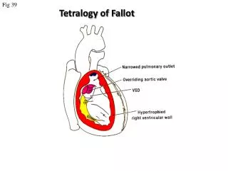
Case presentation- Tetralogy of Fallot - Ventricular Septal Defect. Agatha Stanek. Case presentation. 2 day -old infant presents to ER with mother with severe cyanosis Evident retarded growth, low birth weight Dyspnea upon exertion. Patient hx. Medical Hx

kerry
Download Presentation

Case presentation- Tetralogy of Fallot - Ventricular Septal Defect

An Image/Link below is provided (as is) to download presentation Download Policy: Content on the Website is provided to you AS IS for your information and personal use and may not be sold / licensed / shared on other websites without getting consent from its author. Content is provided to you AS IS for your information and personal use only. Download presentation by click this link. While downloading, if for some reason you are not able to download a presentation, the publisher may have deleted the file from their server. During download, if you can't get a presentation, the file might be deleted by the publisher.

E N D

Presentation Transcript


  1. Case presentation- Tetralogy of Fallot- Ventricular Septal Defect

    Agatha Stanek
  2. Case presentation 2 day -old infant presents to ER with mother with severe cyanosis Evident retarded growth, low birth weight Dyspnea upon exertion
  3. Patient hx Medical Hx Low birth weight; mother gave birth to infant at age of 38 Full-term pregnancy No known allergies yet Family Hx Pseudotruncusarteriosus observed in mother’s niece Some form of cardiovascular diseases, mother reports that grandmother lost 2 children in similar manner- do not know reason why
  4. Social Hx Mother is a lawyer; father is a successful real-estate agent. Live in upper-class suburban area No major stressors in family
  5. Physical Exam Normal arterial and jugular venous pulses Systolic thrill along left sternal border Single S2, decreased P2 HEENT: Retinal engorgement hemoptysis
  6. Differential Diagnosis
  7. Diagnostic procedures Are laboratory tests appropriate? Imaging Tests? Chest X-ray: Doppler echocardiogram:
  8. Diagnostic procedures cont’d Cardiac catheterization Assess severity of right ventricular outflow obstruction Locate position of VSD and its size Rule out possible coronary artery anomalies
  9. Diagnosis Tetralogy of Fallot
  10. Treatment Inpatient care- diagnosis and surgery Surgical measures: Blalock- Taussig shunt or modified shunt (subclavian to pulmnoary artery) Pott’s procedure: descending aorta to pulmonary artery) Waterston’s shunt: ascending aorta to pulmonary artery Total correction surgical therapy- includes patch closure of VSD and relief of right ventricular outflow obstruction
  11. Treatment cont’d. Diet: Salt restriction Activity: As tolerated Medications: No specific drug therapy if there is no heart failure...
  12. Follow-up Patient monitoring: Postoperative Doppler ultrasound 1 year from procedure Post-valvotomy SBE prophylaxis still required Regular follow up assessment for patients not undergoing surgical correction
  13. Possible complications Erythrocytosis? Brain abscess, acute gouty arthritis Infective endocarditis Cerebrovascular thrombosis Postoperative? Fatal if not surgically corrected
More Related