Download
1 / 38
400 likes | 2.01k Views
Radionuclide therapy. External beam radiotherapy. Bevezető. Személyes bemutatkozás(video): Név, iskola bemutatása, néhány kép az iskoláról We are interested in nuclear phisics : We join the Twinning project on THE FIRST EUROPEAN NUCLEAR COMPETITION FOR SECONDARY SCHOOL STUDENTS.

E N D
Radionuclide therapy External beam radiotherapy
Bevezető • Személyes bemutatkozás(video): • Név, iskola bemutatása, néhány kép az iskoláról • Weareinterested in nuclearphisics: • WejointheTwinning project on THE FIRST EUROPEAN NUCLEAR COMPETITION FOR SECONDARY SCHOOL STUDENTS • A nukleáris medicina speciális ágáról a külső sugárkezelésekről (External beam radiotherapy) szeretnénk rövid bemutatót tartani. A projekt készítése előtt rövid látogatást tettünk az Országos Onkológiai Intézetben. • Az ott látottakat is felhasználtuk munkánk során.
What is Nuclear Medicine? • NUCLEAR MEDICINE IMAGINGprocedures look at the bodily functions to help make your diagnosis. • NUCLEAR MEDICINE THERAPYcan actually be used to treat the body. If you are undergoing a therapy process, then larger amounts of radiation will be used to treat cancer or thyroid disease.
What about the radiation? • Very small amounts of radiation are given during nuclear medicine imaging scans. • Larger amounts are used for therapy in order to target very specific areas. • The scanners (equipment) do not give off radiation.
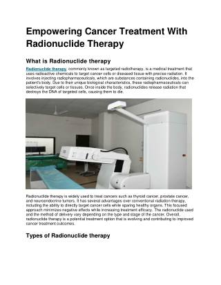
Radionuclide therapy is used in the treatment of both benign disease (eg hyperthyroidism and arthritis) and malignant disease (eg thyroid cancer, Suprarenal gland tumors and hepatocellular carcinoma) Radionuclide therapy
Ourtopic: External beam radiotherapy In the therapeutic issue we must differentiate between • Brachytherapy (internal radiotherapy) sealed source radiotherapy is a form of radiotherapy where a radiation source is placed inside or next to the area requiring treatment. Brachytherapy is commonly used as an effective treatment for cervical, prostate, breast, and skin cancer • and Teletheraby. Kilovoltage ("superficial") X-rays are used for treating skin cancer and superficial structures. Megavoltage ("deep") X-rays are used to treat deep-seated tumors (e.g. bladder, bowel, prostate, lung, or brain).
Gamma-rays, X-rays • There is no consensusfor a definition distinguishing between X-rays and gamma rays. One common practice is to distinguish between the two types of radiation based on their source: X-rays are emitted by electrons, while gamma rays are emitted by the atomic nucleus. This definition has several problems: other processes also can generate these high-energy photons, or sometimes the method of generation is not known. • One common alternative is to distinguish X- and gamma radiation on the basis of wavelength (or, equivalently, frequency or photon energy), with radiation shorter than some arbitrary wavelength, such as 10−11 m (0.1 Å), defined as gamma radiation. This criterion assigns a photon to an unambiguous category, but is only possible if wavelength is known. (Some measurement techniques do not distinguish between detected wavelengths.) However, these two definitions often coincide since the electromagnetic radiation emitted by X-ray tubesgenerally has a longer wavelength and lower photon energy than the radiation emittedby radioactive nuclei. • Occasionally, one term or the other is used in specific contexts due to historical precedent, based on measurement (detection) technique, or based on their intended use rather than their wavelength or source. • Thus, gamma-rays generated for medical and industrial uses, for example radiotherapy, in the ranges of 6–20 MeV, can in this context also be referred to as X-rays.
Beginning: 60Co teletherapy 60Co teletherapy machine for cancer radiotherapy, early 1950s.
Cobaltdecayscheme • Cobalt-60 (60Co), is a synthetic radioactive isotope of cobalt with a half-life of 5.2747 years. It is produced artificially in nuclear reactors. • The activated nickel nucleus emits two gamma rays with energies of 1.17 and 1.33 MeV, hence the overall nuclear equation of the reaction is .
Cobalt decays by beta decay to the stable isotope nickel There is no natural 60Co in existence; thus, synthetic 60Cois created by bombarding a 59Co target with a slow neutron sourceinnuclearreactors.
Kobaltágyú • Külső besugárzásra használt terápiás készülék, melyben a sugárforrás a kobalt radioaktív izotópja (Co-60). A besugárzás a Co-60 által kibocsátott 1,25 MeV átlagenergiájú foton sugárzással történik. A besugárzókészülék fejrészében egy sugárvédett tárolóegységben van a sugárforrás, mely a kezelés elindításakor mechanikus vezérléssel kerül besugárzási pozícióba. A besugárzási idő leteltével a forrás automatikusan visszamegy a tárolóegységbe.
Wearenotabletoregulatethecobaltradiation. What is thesolution? The main advantage of60Co is that it is a high intensity gamma-ray emitter with a relatively long half-life, 5.27 years, compared to other gamma ray sources of similar intensity. The β-decay energy is low and easily shielded; however, the gamma-ray emission lines have energies around 1.3 MeV, and are highly penetrating. Security screening of cars at the Super Bowl using 60Co gamma-ray scanner
Röntgensugarak fajtái keletkezés szerint A széles, folytonos spektrum a fékezési sugárzásból, a vonalszerű spektrum a karakterisztikus sugárzásból származik. A fékezési sugárzást a nagy rendszámú atommagok erős elektromos terén szóródó elektronok hozzák létre. A lefékeződés során az elektronok energiájuk kis részét röntgenfotonok formájában kisugározzák, az energia másik része pedig hővé alakul. A sugárzás spektruma folytonos, a rövid hullámhosszú oldalon éles határral. A karakterisztikus sugárzás úgy jön létre, hogy az anódba becsapódó elég nagy energiájú elektron képes az atom egy az atommaghoz közeli, belső elektronhéjon lévő elektronját kiütni. Az így megüresedő energiaszintű állapotra aztán egy magasabb energiájú elektron kerül, és az átmenet során az energiakülönbségnek megfelelő röntgenfoton emittálódik. Spektruma vonalas, a vonalak helyzete az adott atomra jellemző.
Generating gamma rays History: X-rays make up X-radiation, a form of electromagnetic radiation. Most X-rays have a wavelengthranging from 0.01 to 10 nanometers, corresponding to frequencies in the range 30 petahertz to 30 exahertz (3×10¹⁶ Hz to 3×10¹⁹ Hz) and energies in the range 100 eV to 100 keV. X-ray wavelengths are shorter than those of UV rays and typically longer than those of gamma rays. In many languages, X-radiation is referred to with terms meaning Röntgen radiation, after the German scientist Wilhelm Röntgen who discovered these on November 8, 1895, who usually is credited as its discoverer, and who named it X-radiation to signify an unknown type of radiation.]Spelling of X-ray(s) in the English language includes the variants x-ray(s), xray(s), and X ray(s) The gamma-rays aregenerated for medical and industrial uses, for example radiotherapy, in the ranges of 6–20 MeV.
Characteristic X-ray emission • Characteristic X-ray emission (X-ray fluorescence): If the electron has enough energy it can knock an orbital electron out of the inner electron shell of a metal atom, and as a result electrons from higher energy levels then fill up the vacancy and X-ray photons are emitted. This process produces an emission spectrum of X-rays at a few discrete frequencies, sometimes referred to as the spectral lines. The spectral lines generated depend on the target (anode) element used and thus are called characteristic lines. Usually these are transitions from upper shells into K shell (called K lins), into L shell (called L lines) and so on
Bremsstrahlung • This is radiation given off by the electrons as they are scattered by the strong electric field near the high-Z (proton number) nuclei. These X-rays have a continuous spectrum. The intensity of the X-rays increases linearly with decreasing frequency, from zero at the energy of the incident electrons, the voltage on the X-ray tube.
Bremsstrahlung (braking radiation) • Bremsstrahlung (German), from bremsen "to brake" and Strahlung "radiation"; i.e., "braking radiation" or "deceleration radiation", is electromagnetic radiation produced by the deceleration of a charged particle when deflected by another charged particle, typically an electron by an atomic nucleus. • The moving particle loses kinetic energy, which is converted into radiation (i.e., a photon), thus satisfying the law of conservation of energy. • The term is also used to refer to the process of producing the radiation. Bremsstrahlung has a continuous spectrum, which becomes more intense and whose peak intensity shifts toward higher frequencies as the change of the energy of the decelerated particles increases
X-rays are created by two different atomic processes Spectrum of the X-rays emitted by an X-ray tube with a rhodium target, operated at 60 kV. The smooth, continuous curve is due to bremsstrahlung, and the spikes are characteristic K lines for rhodium atoms.
Linear accelerator (Linac) • Animation showing how a linear accelerator works. In this example the particles accelerated (red dots) are assumed to have a positive charge. The animation shows a single particle being accelerated each cycle; in actual linacs a large number of particles are injected and accelerated each cycle. The graph V(x) shows the electrical potential along the axis of the accelerator at each point in time. The polarity of the RF voltage reverses as the particle passes through each electrode, so when the particle crosses each gap the electric field (E, arrows) has the correct direction to accelerate it. The action is shown slowed down enormously.
The first patient treated in 1953 in London • Linac-based radiation therapy for cancer treatment began with the first patient treated in 1953 in London, UK, at the Hammersmith Hospital, with an 8 MV machine built by Metropolitan-Vickers and installed in 1952, as the first dedicated medical linac. A short while later in 1954, a 6 MV linac was installed in Stanford, USA, which began treatments in 1956.
Visiting (National Institute of Oncology ) Equipmentsforexternalbeamradiotherapy 5 linearaccelerators (LINACs): • Varian TrueBeam 6, 10 & 18 MV photons, 6-18 MeVelectrons, image-guided (IGRT) and intensitymodulatedradiotherapy (IMRT), gatedradiotherapy, stereotacticradiosurgery (SRS) and sterotactic body radiotherapy (SBRT) • Siemens Artiste 6 & 18 MV photons, 6-18 MeVelectrons, IGRT, IMRT, gatedradiotherapy • Siemens Primus I 6 & 18 MV photons, 6-21 MeVelectrons, 3D conformalradiotherapy (3D-CRT) • Siemens Primus II 6 & 18 MV photons, 6-21 MeVelectrons, 3D-CRT, IMRT, SRS, micromultileaf collimator (micro-MLC) • Siemens Primus III 6 MV photon, 3D-CRT Alllinearacceleratorsareequipedwithmultileaf collimator (MLC),and electronicportalimagingdevice (EPID). The Varian TrueBeammachine is mountedwith a kV cone-beamCT,whileour Siemens Artiste LINAC is using MV cone-beam CT and an „in-room” CT unit forhighprecision IGRT and IMRT.
CyberKnife CyberKnife is a highly conformal radiosurgical technology which can successfully treat this subset of patients. In addition, it can be applied for hardly resectable rare tumors of the skull base and the head and neck region like chordoma, chondrosarcoma and paragangliomas. The
ARTISTE, an Integrated Dose-Guided Radiation Therapy Solution This advanced system for the delivery of ART will provide both kilovoltage (kV) and megavoltage (MV) imaging capabilities by providing a separate radiation source and imaging panel for each energy range. Each energy range is then used to provide the images necessary for DGRT, and can become the basis of distinct treatment and imaging capabilities. The arrangement of the sources and imaging panels are 180 degrees (in-line) from one another, which allows ARTISTE to be unique in the intent to provide the only solution able to image both the patient and the treatment at the same time. In addition, ARTISTE will provide synchronized image and dose monitoring, quality cone beam information correlated with the treatment plan, and both entrance and exit treatment ports.
TrueBeamHigh-PrecisionRadiotherapyTreatment • TrueBeam™ redefines radiotherapy as we know it. Faster to administer than conventional Radiotherapy, TrueBeamrotates around the patient to deliver powerful, specifically targeted beams of radiation with pinpoint accuracy. • The system’s power and flexibility allows clinicians to develop personalized treatments for patients’ individual circumstances. • TrueBeam can be used to treat a diverse range of cancers, including tumors of the lung, breast, head and neck, abdomen, liver, and other regions.
Precision • The precision of Varian’s TrueBeam system is measured in increments of less than a millimeter. The system performs accuracy checks every ten milliseconds. • This helps the radiation oncologist protect critical organs and healthy tissue surrounding the tumor. • The system design also gives the treatment team more options for positioning the patient. • In addition, TrueBeam has a new “gated” option to synchronize beam delivery with the patient’s breathing. • This helps maintain accuracy in situations where tumor motion is an issue, such as during lung cancer treatments.
Speed. Efficiency Speed With TrueBeam, treatments that once took 10 to 30 minutes can now be completed in less than two minutes. With faster treatment, there is less chance of tumor and patient movement. What’s more, the treatment is more comfortable and less stressful for patients. Efficiency TrueBeam’s sophisticated open architecture integrates imaging, real-time tracking, respiratory gating, and treatment delivery. This makes it possible to deliver treatments more quickly and accurately, while monitoring and adjusting for tumor motion. TrueBeam rotates around the patient to deliver a prescribed radiation dose from nearly any angle.
TrueBeam • A készülék különböző energiájú foton- és elektronsugárzás előállítására képes, és speciális tulajdonsága a nagy intenzitású sugárnyalábbal történő kezelés, amellyel jelentősen lerövidül a besugárzási idő. A berendezés fontos része az integrált képalkotás, ami röntgenfelvételekkel és CT vizsgálattal teszi lehetővé az ún. képvezérelt sugárterápiát. Ennek lényege, hogy a sugárkezelések megkezdése előtt közvetlenül, vagy akár közben is, képalkotó eljárásokkal ellenőrzik és javítják a betegbeállítás pontosságát. Ezt segíti a robotikus kezelőasztal, ami minden irányban távvezérléssel mozgatható és forgatható. A módszer előnye az ép szövetek és a védendő szervek nagyobb fokú kímélete, ami a mellékhatások csökkenéséhez vezet. A TrueBeam készülék lehetővé teszi az ún. forgóíves intenzitásmodulált sugárkezelést is, mellyel szabálytalan alakú daganatok besugárzása rövid idő alatt úgy végezhető el, hogy a közelben levő egyéb szervek és egészséges szövetek dózisterhelése minimális marad. A készülék alkalmas mozgó céltérfogatok besugárzására is, elsősorban a mellkasi régióban a légzés miatti daganatelmozdulásvalósidejű követésével. Nagyfokú geometriai pontosság és a képi ellenőrzés miatt, kisméretű daganatok sztereotaxiás technikával történő besugárzása is végezhető vele koponyán belüli és azon kívüli régiókban is. • A TrueBeam lineáris gyorsító jelenleg az egyik legújabb és legmodernebb onkológiai besugárzó készülék a világon, mellyel a legnevesebb amerikai és nyugat-európai onkológiai központok is dolgoznak. A Közép-európai régióban ez az egyetlen ilyen típusú készülék. Használatával az OOI újabb nemzetközi klinikai kutatási programokba kapcsolódhat be.
Source • https://en.wikipedia.org/wiki/X-ray • https://www.youtube.com/watch?v=jSgnWfbEx1A • https://en.wikipedia.org/wiki/Bremsstrahlung • https://www.flinnsci.com/flinn-electromagnetic-spectrum-chart/ap7148/ • https://en.wikipedia.org/wiki/Linear_particle_accelerator • https://www.google.hu/search?rlz=1C1WSCA_enHU569HU569&q=cobalt+decay+scheme&tbm=isch&source=univ&sa=X&ved=2ahUKEwiiiuL9v_jgAhVx-ioKHcBIC_wQsAR6BAgCEAE&biw=1280&bih=640#imgrc=NTzRM4qQzPejaM: • https://en.wikipedia.org/wiki/Cobalt-60#/media/File:Nci-vol-1819-300_cobalt_60_therapy.jpg • https://en.wikipedia.org/wiki/Cobalt-60 • http://www.onkol.hu/sites/default/files/osztalyok/sugarterapias_osztaly_keszulekek_2014.pdf • https://www.oncologysystems.com/inventory/medical-equipment-for-sale/siemens-artiste-linear-accelerator • http://www.patikamagazin.hu/a-legmodernebb-besugarzo-keszulek-az-orszagos-onkologiai-intezetben/