Download
1 / 38
380 likes | 559 Views
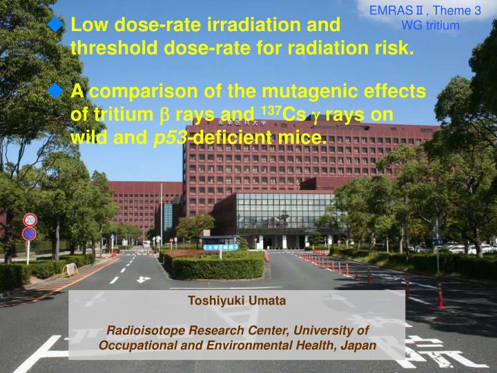
Low dose-rate irradiation and threshold dose-rate for radiation risk. A comparison of the mutagenic effects of tritium rays and 137 Cs rays on wild and p53- deficient mice. EMRAS Ⅱ, Theme 3 WG tritium. Toshiyuki Umata Radioisotope Research Center, University of

E N D
Low dose-rate irradiation and threshold dose-rate for radiation risk. • A comparison of the mutagenic effects of tritium rays and 137Cs rays on wild and p53-deficient mice. EMRASⅡ, Theme 3 WG tritium Toshiyuki Umata Radioisotope Research Center, University of Occupational and Environmental Health, Japan
Low dose-rate irradiation and threshold dose-rate for radiation risk. • A comparison of the mutagenic effects of tritium rays and 137Cs rays on wild and p53-deficient mice.
OBJECTIVE To investigate the biological effects of tritium on mouse at low dose-rate, mice were exposed to -rays by continuous administration of various concentration of tritiated drinking water throughout their lives at low dose-rates. CONCLUSION These studies revealed that there exists two types of threshold dose-rates, not only in the frequency of thymic lymphomas but also in the life-shortening. In the life-shortening, it seems that the effect of tritium rays is greater than that of rays.
7 blood organ tissue (average of brain, Liver, muscle)
Table1 Tumor developments in mice at different dose-rates of HTO Dose-rate, Gy/day 0.240.0 960.0480.024 0.010 0 Number of mice used 45 38 60 60 53 67 MST, days 165±36259±52414±66481±112622±121811±134 Thymic lymphoma 29(64)[162±28]22(58)[273±51] 15(25)[415±53]4(7)[508±202] 3(6)[589±32] 0(0) Non-thymic lymphoma 5(11)[146±27]4(11)[229±24] 12(20)[433±82] 9(15)[504±120] 11(21)[609±70] 12(18)[787±129] Reticular cell neoplasm 2(5)[179±15] 5(8)[390±67] 12(20)[485±144] 10(19)[570±150] 4(6)[760±161] Ovarian tumor2(5)[201±18] 4(7)[431±60] 8(13)[511±98] 11(21)[641±114] 4(6)[868±149] Haemangiosarcoma2(5)[331±21] Fibrosarcoma 2(3)[431±58] 4(7)[467±97] 6(11)[607±90] 4(6)[871±179] Harderian gland tumor2(3)[423±81] 2(3)[537±75] Lung tumor 1(2)[464] 3(5)[460±30] 8(15)[736±84] 4(6)[812±24] Skin tumor 1(2)[401] Bladder tumor1(2)[580] Rhabdomyosarcoma1(2)[298] Mammary tumor2(4)[582±58] Hepatic tumor2(4)[685±23] 3(4)[696±41] Adenal gland tumor1(2)[623] Splenic tumor2(3)[827±19] Stomac tumor1(2)[912] Double tumor-bearing 0(0) 0(0) 0(0) 2(3) 10(19) 5(8) Tumor-bearing mouse 34(76) 32(84) 42(70) 42(70) 4(83) 41(54) ( );% [ ];Mean latent period ±SD, day MST: mean survival time or mean time of death after the initiation of the exposure.
Table2 Tumor developments in mice at different dose-rates of HTO Dose-rate, mGy/dayControl 1*Control 2Total control0.2 0.9 3.6 (Cumulative dose, Gy) (0) (0) (0) (0.17±0.03) (0.71±0.13) (2.62±0.41) Number of mice69 51 120 55 58 120 MST, days810795804790758804 Thymic lymphoma 0(0) 0(0) 0(0) 0(0) 0(0) 3(5)[556±40] Non-thymic lymphoma 16(23)[773±147]14(27)[799±113] 30(25)[787±129] 14(25)[769±137] 17(29)[779±84] 11(20)[728±97] Fibrosarcoma5(7)[860±161] 4(8)[81±126] 9(8)[838±145] 5(9)[836±36] 13(22)[767±116] 5(9)[835±18] Ovarian tumor4(6)[868±149] 2(4)[885±80] 6(5)[875±121] 3(5)[829±187] 2(3)[811±197] 5(9)[819±111] Livertumor6(9)[800±151] 3(6)[729±202] 9(8)[776±168] 4(7)[939±62] 10(17)[844±120] 1(2)[729] Lung tumor3(4)[811±24] 2(4)[967±22] 5(4)[873±23] 4(7)[847±115] 1(2)[852] Other tumors 3(4)[855±40] 3(6)[775±139] 6(5)[815±110] 4(7)[920±78] 3(5)[879±91] 2(4)[941±3] Multiple tumor-bearing 4(6) 2(4) 6(5) 3(5) 4(7) 2(4) Tumor-carrying mice 33(48) 26(51) 59(49) 27(49) 45(78) 26(46) ( );% [ ];Mean latent period ±SD, day MST: mean survival time or mean time of death after the initiation of the exposure. *Previously reported (Yamamoto et al. 1995)
20 Gy 400 days 25% 2×10⁹ Bq/L 50 mGy/day =18 Gy/y
SUMMARY • There exists two types of threshold dose-rates, essential and practical, not only in the frequency of thymic lymphoma but also in the life-shortening. In the life-shortening, it seems that the effect of tritium rays is greater than that of rays. • Threshold dose-ratein the frequency of thymic lymphomas was12 (0.9) mGy/day. And that in the life-shortening was 2 (0.2) mGy/day (1.8 Gy).
Low dose-rate irradiation and threshold dose-rate for radiation risk. • A comparison of the mutagenic effects of tritium rays and 137Cs rays on wild and p53-deficient mice.
OBJECTIVES Although nuclear fusion facilities, such as ITER, are expected to require about 2,000 PBq of tritium for their “fuel”, only a small part of these tritium may be released from the facilities. Therefore, an exposure condition of tritium radiation from nuclear fusion reactor could be a long-term exposure at a low dose-rate. So, analytical methods sensitive to radiation at low dose or low dose-rate are needed. In the present study, the mutagenic effects of tritiated water at a low dose-rate was investigated using wild type and p53-/-mice, then compared that of 137Cs rays.
Features of p53-/- mice ・ p53 is a tumor suppressorgene. ・ p53 plays the role as guardian of genome. ・ p53-/- mice have no apoptotic activity, which remove damaged cells from tissue. ・ Life of p53-/- mice are short cause of occurrence of tumor. : ~ 8 months (wilt-type mice: ~28 months)
Preparation of T lymphocytes for Mutation Analysis hemolysis of red cells gently dissociate 37℃1 h spleens filtration nylon wool cell suspension ・PE conjugated anti-CD4 antibody ・FITC conjugated anti-CD3 antibody T-lymphocytes FACS 5 x 10⁵ cells
TCR mutation assay (principle) CD3 CD4 T cell receptor Extracellular Cytoplasm irradiation Extracellular Cytoplasm If mutation is formed in T cell receptor, CD3 is not able to be transfered into cell membrane. Mut Res 1997, Meth Mol Biol 2005, Radiat Res 2006
0 Gy (control mouse) 3 Gy (irradiated mouse) CD3+CD4+ CD3+CD4+ CD3-CD4+ CD3-CD4+ PE-anti CD4 PE-anti CD4 FITC-anti CD3 FITC-anti CD3 CD3-CD4+ CD3-CD4+ TCR VF = TCR VF = CD3+CD4+ CD3+CD4+ 37 188 = =7.4×10-4 = = 37.5×10⁻⁴ 50191 50097 Estimation of TCR variant fraction (mutation frequency)
Irradiation methods Intraperitoneal injection of HTO (266 MBq/mouse) and start of simulation-irradiation (0.7 mGy/min at start) Acute irradiation(3Gy) 0.8 4 absorbed dose 0.7 Dose rate (mGy/min) 3 0.6 Cumulative absorbed dose (Gy) 0.5 0.4 2 0.3 dose rate 0.2 1 0.1 0 0 9 10 11 12 13 14 15 16 17 18 19 Days after the start of irradiation Days after the start of irradiation Analysis of mutation [p53+/+, p53-/-] 0 1 2 3 4 5 6 7 8 Analysis of apoptosis [ p53+/+, p53-/-] 8 week old normal and p53-/- mice were used. Mice were irradiated by 137Cs -rays simulation-irradiation system in 2.3 days as an effective half-life.
137Cs g-rays simulation- irradiation system for low dose-rate
:spontaneous :HTO :137Cs γ- rays (<0.7 mGy/min) :137Cs γ- rays (0.86 Gy/min) 40 3 Gy 35 30 * P < 0.01 * * 25 Variant fraction (x10-4) 20 * 15 10 5 0 p53+/+mice p53-/- mice
Induced TCR variant fractions Relative value induced variant fraction mice exposure A / B A / C - A 3.9 x 10⁻⁴ HTO 0.3 - - p53+/+ 0 simulation B - 14.2 x 10⁻⁴ 1.0 acute g-rays C 7.6 x 10⁻⁴ HTO 0.4 1.7 A - 4.5 x 10⁻⁴ 1.0 simulation B p53-/- 20.1 x 10⁻⁴ - 1.0 acute g-rays C
A D A E B F C p53+/+ p53-/- spontaneous HTO 137Cs
SUMMARY • To evaluate the mutagenic effects of tritiated water, TCR variant fractions using wild type and p53-/- mice were investigated. • When compared on the basis of the induced TCR variant fractions in p53 deficient mice at 3 Gy, tritium rays appear to be 1.7 times more mutagenic han rays. • p53-dependent apoptosis could suppress the increase of TCR variant fraction induced by tritium rays.
Radioisotope Research Center Thank you for your attention
Irradiation methods Intraperitoneal injection of HTO (266 MBq/mouse) and start of simulation-irradiation (0.7 mGy/min at start) Acute irradiation(3Gy) 0.8 4 absorbed dose 0.7 Dose rate (mGy/min) 3 0.6 Cumulative absorbed dose (Gy) 0.5 0.4 2 0.3 dose rate 0.2 1 0.1 0 0 9 10 11 12 13 14 15 16 17 18 19 Days after the start of irradiation Days after the start of irradiation Analysis of mutation [p53+/+, p53-/-] 0 1 2 3 4 5 6 7 8 Analysis of apoptosis [ p53+/+, p53-/-] 8 week old normal and p53-/- mice were used. Mice were irradiated by 137Cs -rays simulation-irradiation system in 2.3 days as an effective half-life.
Induced TCR variant fractions Relative value induced variant fraction mice exposure A / B A / C - A 3.9 x 10⁻⁴ HTO 0.3 - - p53+/+ 0 simulation B - 14.2 x 10⁻⁴ 1.0 acute g-rays C 7.6 x 10⁻⁴ HTO 0.4 1.7 A - 4.5 x 10⁻⁴ 1.0 simulation B p53-/- 20.1 x 10⁻⁴ - 1.0 acute g-rays C
A D A E B F C p53+/+ p53-/- spontaneous HTO 137Cs
Researches for biological effect of HTO on mice End Point administration dose(Gy) control RBE reference LD50/30 i.p4~81.7 Furchner Radiat Res 1957 Contraction of spleen i.p 1 ~10 1.3 - 1.5 Storer et al. Radiat Res and Thymus 1957 genomic instability of 0.6 1 - 2 Kozkowski et al. lymphocyte Int J Radiat Biol 2001 apoptosis of crypt cell0.13~0.28 1.4 - 2.1Ijiri Radiat Res 1989 survival of oocyte0.055 1.6 - 3 Dobson et al. Radiat Res 1976 i.p. : intraperitoneally ・対照放射線
principle The T lymphocyte is one of the most radiosensitive cells in the body. Several genetic end points are used to measure mutant fractions in blood cells. The T-cell receptor (TCR) gene is utilized for in vivo and in vitro mutation assays in both human and animal cells. It is known that the TCR-α and TCR-βchains are only expressed on the cell surface following the formation of large molecular complexes with CD3 chains. If either the TCR-αor -βchain gene is not expressed, the TCRαβ/CD3 complex cannot be transported to the cellular membrane, resulting in the accumulation of defective complexes in the cytoplasm. Thus, inactivation mutations ineither the TCR-α or TCR-β genes in CD4+ T cells manifest as CD3-CD4+ variant T cells, that can be detected via two-color flow cytometry using monoclonal antibodies against the CD3 and CD4 molecules. In the present study, the TCR assay was utilized to quantify the genotoxic effects of tritium exposure in mice.
∞ TableTissue cumulative absorbed dose by HTO (Gy) •18.5MBq HTO were i.p. injected 7日間で3Gyの吸収線量を達成するには 3/0.208X 18.5 =266 MBq を接種する。 J UOEH 1988
低線量でのがん発生の線量ー効果モデル Dose-effect model 実測域 LNT hypothesis 推定域 Frequency of Tumor (%) a0 spontaneous (low dose) (high dose) 0 Dose
p53 (+/+) p53 (–/–) repair repair error repair repair error apoptosis removal of abnormal cell abnormal cell normal cell normal cell 3 Gy 3 Gy Variant fraction High Dose-rate High Dose-rate Low Dose-rate Low Dose-rate Background level Background level p53 (–/–) p53 (+/+) p53 System against Genotoxic Stress
50 p53–/– 50 @ 1.2 mGy/min (@ 1,017 mGy/min) 40 40 Variant fraction (x 10 –4) 30 Variant fraction (x 10 –4) 30 p53 –/– p53+/+ 20 20 p53+/+ 10 10 0 0 1 2 0 3 1 2 3 0 Dose (Gy) Dose (Gy) Dose response curves at different dose-rate of g rays for the TCR variant fractions in the splenic T-lymphocytes of p53+/+ and p53–/– mice Kato et al, J Radiat Res 2002
OBJECTIVE There are a number of important variables that may alter the precise estimation of the radiation weighting factor (WR) for tritium b rays. For instance, the interpretation of published experimental studies is complicated by the fact that the dose rates of reference radiations are frequently much higher than those normally received by tritium internal exposure. To resolve these uncertainties and determine the most appropriate WR for tritium b rays, the mutagenic effects of tritium b rays on spleen T lymphocytes of Trp53+/+ and Trp53-/- mice was compared to chronic or acute 137Cs g irradiation.