Download
1 / 39
390 likes | 494 Views
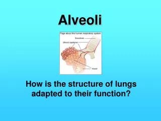
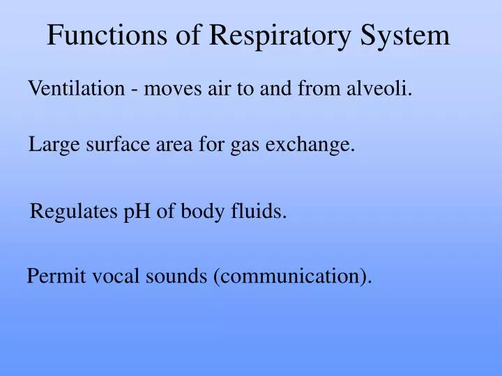
Functions of Respiratory System . Ventilation - moves air to and from alveoli. Large surface area for gas exchange. Regulates pH of body fluids. Permit vocal sounds (communication). External nares. Upper Respiratory Tract Nose Nasal cavity Pharynx (3 parts). Conditions inspired air:.

E N D
Functions of Respiratory System Ventilation - moves air to and from alveoli. Large surface area for gas exchange. Regulates pH of body fluids. Permit vocal sounds (communication).
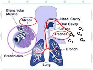
Upper Respiratory Tract • Nose • Nasal cavity • Pharynx (3 parts) Conditions inspired air: Filter Warm Humidify • Lower Respiratory Tract - Larynx - Trachea - Bronchi - Bronchioles - Alveoli Functions to conduct air to site of gas exchange.
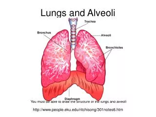
Respiratory Tract (Passageway) External nares > nose > nasal cavity > pharynx (naso-, oro- laryngo-) > larynx > trachea > 1o bronchi > 2o bronchi > 3o bronchi > bronchioles > terminal bronchioles > respiratory bronchioles > alveolar duct > alveolar sac > alveoli (end).
The Respiratory Tract is divided into: • Conducting Zone • Respiratory Zone
Two types of mucosa in nasal cavity • Olfactory mucosa – houses olfactory receptors • Respiratory mucosa – lines nasal cavity PARANASAL SINUSES
“Respiratory Epithelium” • Lines conducting portions of tract. • Pseudostratified ciliated columnar epithelium (with goblet cells) • Produces mucus to trap foreign particles • Lamina propria is the connective tissue layer (Epithelium and lamina propria = mucus membrane)
External nares • Open into nasal cavity • Vestibule guarded by hairs • Nasal cavity • Superior, middle and inferior meatuses • Narrow grooves and conchal surfaces • Hard palate • Divides nasal and oral cavities • Soft palate • Superior nasopharynx and pharynx • Internal nares • Between nasal cavity and nasopharynx
The Pharynx Portions shared by digestive and respiratory systems Nasopharynx Superior portion: from internal nares to uvula. Oropharynx Continuous with oral cavity Laryngopharynx Between hyoid and entrance to esophagus
Nasopharynx • Superior to where food enters. • Only an air passageway. • Closed off during swallowing (uvula). • Pharyngeal tonsil (adenoids) • Located on posterior wall • Destroys entering pathogens • Contains the opening to the auditory tube
Oropharynx • Extends from soft palate to the epiglottis. • Stratified squamous epithelium. • Two types of tonsils in the oropharynx Palatine tonsils – in the lateral walls of fauces. Lingual tonsils – on posterior surface of tongue.
Laryngopharynx • Passageway for both food and air. (shared respiratory and digestive) • Stratified squamous epithelium • Continuous with the esophagus and larynx
Lower Respiratory Tract: The Larynx • Surrounds glottis - air passes through glottis to reach lungs • Epiglottis - prevents solids from entering respiratory system Laryngeal cartilages
When swallowing, elevation of larynx folds epiglottis over glottis, steering materials into the esophagus.
Trachea • From C6 to T5 • About 4.5 inches in length. • About 1 inch in diameter. • Submucosa includes “C” rings of cartilage Posterior wall (without cartilage) distorts, allowing food passage through esophagus. • Contains the muscle trachialis.
Trachea 1) pseudostratified ciliated columnar epi2) tracheal cartilage ring3) trachealis muscle 1) 2) 3)
Left and Right 1o Bronchi • Right and left primary bronchi • Trachea branches within mediastinum. • Bronchial tree • Enters lungs at hilus (“entry/exit”). – Extensively branching passageways.
Lungs • Separated by fissures • Right lung has three lobes. • Left lung has two lobes. • Costal surface • Anterior surface • Follows inner contours of rib cage • Mediastinal surface • Contains hilus • Left lung bears costal notch
For clarity, the degree of branching has been reduced: an airway branches approximately 23 times before reaching the level of a lobule.
Basic structure of a lobule, cut to reveal the arrangement between the alveolar ducts and alveoli.
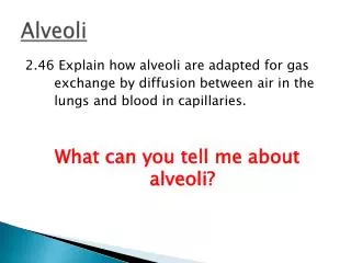
Alveoli consist of 3 types of cells 1)Alveolar Type I cells – simple squamous epithelium, make the ‘walls’ of alveoli for gas exchange. 2)Alveolar Type II cells – simple cuboidal cells, release surfactant, help make lungs Compliant. 3)Alveolar Macrophages – phagocytic cells, protects the alveolar surface.
Respiratory Muscles Ventilation - movement of air into and out of lungs.
Eupnea - normal quite breathing at rest. Inspiration: volume of thoracic cavity. Muscle activity required: Diaphragm External Intercostals Sternocleidomastoid Scalenes
To increase depth and frequency of breaths: Sternocleidomastiod Scalenes Expiration: volume of thoracic cavity. No Muscular activity required (in eupnea)
When Forcefully exhaling (hypereupnea): Muscles used: Internal Intercostals Rectus abdominis Transverse abdominis, Internal and External obliques.
Positions and relationships between the major respiratory centers.
Three pairs of nuclei in reticular formation of pons and medulla oblongata Respiratory rhythmicity center - Sets respiratory pace. Located in the medulla oblongata. Dorsal Respiratory Group (DPG) = inspiration. Ventral Respiratory Group (VPG) = forced breathing. Apneustic center - Strong, sustained inspiratory movements, used for ‘overdrive’ when breathing deep. Pons Pneumotaxic center - Inhibits apneustic and inspiratory centers, limits over inflation of lungs. Pons
Sensory Receptors - regulate respiration. Mechanoreceptors detect changes in lung volume or arterial blood pressure Chemoreceptors Changes in PCO2, pH, PO2 of blood andCSF Central chemoreceptors - in medulla Peripheral chemoreceptors Aortic bodies (in aorta) Carotid bodies (in carotids)