Download
1 / 59
640 likes | 1.05k Views
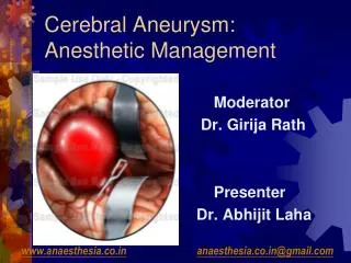
ANEURYSM Clinical presentation, Diagnosis and Anaesthetic management. Dr. Girija Rath Dr. Divya Dr. Abhijit. www.anaesthesia.co.in anaesthesia.co.in@gmail.com. Intracranial Aneurysms. Most common cause of subarachnoid haemorrhage Types - Saccular, Fusiform, Dissecting

E N D
ANEURYSMClinical presentation, Diagnosis and Anaesthetic management Dr. Girija Rath Dr. Divya Dr. Abhijit www.anaesthesia.co.inanaesthesia.co.in@gmail.com
Intracranial Aneurysms • Most common cause of subarachnoid haemorrhage • Types - Saccular, Fusiform, Dissecting • Size – Small <12 mm in diameter (78%) Large -12-24 mm (20%) or Giant >24 mm (2%) • 50 – 80% don't rupture
Epidemiology • Prevalence of Intra cranial aneurysms – 1-5% adult population • Incidence of SAH from rupture of Intra cranial aneurysms - 1 / 10,000 persons • Ruptured Aneurysm - 75- 85% case of SAH
Epidemiology (cont.) • Risk of SAH - 2 times more in woman • Peak incidence in 55- 60 yrs • SAH – high mortality and morbidity (2 of 3 survive to receive medical t/t; 1 of 3 receiving t/t will be severely disabled or die)
Associated conditions • Mostly sporadic lesions • Rare familial form has also been described • Associated conditions – autosomal dominant polycystic kidney disease, fibromuscular dysplasia, Ehler – danlos syndrome , AV malformations of brain, coactation of aorta • Smoking and hypertension - ↑ risk • Vascular abn. – Type 3 collagen deficiency
Pathogenesis • Acquired vascular lesions secondary to degenerative changes in vessel wall • Hypertension and smoking induced vascular changes – major predisposing role • Common histological finding - ↓ tunica media in middle muscular layer of artery
Distribution • occur at vascular branching points • most centered around the circle of Willis • 20-30% are multiple, and often mirror • usually rupture at the dome
Distribution Anterior circulation (85%) • ACA complex incl. Anterior Communicating (AComm) & pericallosal artery • MCA bi-/trifurcation • Internal Carotid Artery (esp at origin of PComm, ophthalmic, anterior choroidal, or terminus) Posterior Circulation (15%) • basilar tip • also PICA
Clinical presentation of Aneurysm • Most common presentation - Subarachnoid haemorrhage • Silent - incidental finding on investigations • Symptomatic – d/t mass effect cranial nerve palasies ( 3 nerve palasy in PCA aneurysm) brain stem compression
Natural History of Aneurysm • Ruptured aneurysms – greater chance of future rupture • Unruptured aneurysms - 1-2 % per year risk of rupture Exceptions • symptomatic aneurysms, size > 10 mm, • aneurysms of basilar apex or PCA • Coexisting or remaining aneurysms of all sizes in pts with H/O SAH ( AHA Recommendations for the Management of unruptured Intracranial Aneurysms – 2001)
Pathophysiology of Aneurysm Rupture • Rupture aneurysm initial increase in ICP decrease in CBF and CPP immediate change in the level of consciousness • The dramatic reduction of CBF is a major factor halting the subarachnoid bleeding.
Pathophysiology of Aneurysm Rupture (cont.) Two distinct cerebral hemodynamic patterns are seen • Gradual reduction in ICP increase in CBF over approx.15 min improvement of cerebral function. Clinically patients present with varying levels of consciousness. • Persistent increase in ICP resulting in failure of recovery of both CBF and functional activity. Clinically these pts. die before receiving treatment or have persistent vegetative state.
Clinical Features of Ruptured Aneurysm/SAH Neurological • Headache - severe ("worst in my life!") • often transient loss of consciousnessat onset (due to abrupt rise in ICP) • Signs of meningeal irritation – neck stiffness • Nausea & Vomiting, photophobia, altered mentation • Focal Deficits – sensory, motor, visual
Clinical Features of SAH (cont.) Cardiovascular - ECG changes – 50 to 80% • Prolongation of the Q-T interval, ST and T wave changes, P wave changes, U waves, and dysrythmias including VT & VF • Usually during first 48 hrs. Normalise in 10 days • Cause mostly Neurogenic – SAH related injury to the posterior hypothalamus stimulate release of norepinephrine from the adrenal medulla and sympathetic cardiac efferent (Stroke 1991;22:746-9.) • 27 -33% - ventricular wall dysfunction • D/D - electrolyte disturbances, Myocardial ischemia/ infarction
World Federation of Neurological Surgeon’sGrading Scale - WFNS GradeGCSMotor Deficit • I 15 Absent • II 13-14 Absent • III 13-14 Present • IV 7-12 +/- • V 3-6 +/-
Diagnostic Studies • Non contrast CT • Lumbar puncture • MRI • Angiography
Noncontrast CT • Procedure of choice for confirming diagnosis of SAH • CT - magnitute and location of SAH probable location of aneurysm ventricular size • Prospective series of 100 patients with suspected SAH 100% within 48 hrs 85% after 5 days 50% after 1 week 30% after 2 weeks Van Gijn et al. Time course of aneurysmal hemorrhage on CT Neuroradiology 23 : 1985
Fisher Grading Base on the amt and distribution of SAH blood on NCCT Predicts the probability of developing delayed ischaemia d/t vasospasm • 1 No subarachnoid blood detected • 2 Diffuse or vertical layers< 1 mm thick • 3 Localized clot and/or vertical layer > 1 mm • 4 Intracerebral or intraventricular clot with diffuse or no subarachnoid blood
Lumbar puncture • Before advent of CT, only modality available • Reserved for 5% patients with negative CT • False positive d/t traumatic tap • xanthochromia (yellow color) is more sensitive sign than bloody CSF (appears after 4 hrs , negative after 3 weeks) • Dangerous in patients with raised ICP - risk of brain herniation and aneurysm rebleeding
MRI • Magnetic resonance imaging is not well suited for imaging SAH in the acute stage • After several days or weeks, when the CT scan has normalized, MRI may detect subpial hemosiderin close to the source of the rupture.
Cerebral angiography • Gold standard for identifying aneursyms • 4 vessel angiogram should be performed as soon as possible after the diagnosis of SAH to identify etiology (aneursym rupture 75 -85%) • Presence of multiple aneurysm (incidence 5%-33.5%)
CT angiography • noninvasive modality • In aneurysms > 3 mm sensitivity 90 – 95% specificity 100% Harrison MJ et al. neurosurg 40 : 1997
MRA – Magnetic resonance angiography • Probably the best noninvasive modality to detect etiology of SAH • In aneurysms > 3 mm sensitivity 91 – 96% specificity 100% (Schwartz RB et al. radiology 192 : 1994) • More useful in patients with angio negative SAH renal failure contrast c/i
Screening for Asymptomatic Aneurysms Screening with MRA is indicated • for people who have two immediate relatives with Intra cranial aneurysm • for all patients with autosomal dominant polycystic kidney disease An estimated 5 to 40 percent of patients with autosomal dominant polycystic kidney disease have intracranial aneurysms
Complications of Ruptured aneurysm/SAH • Symptomatic vasospasm • Rebleeding • Hydrocephalus • Seizures • Hyponatremia • The leading causes of death and disability after SAH are direct effect of the initial bleed, cerebral vasospasm, and rebleeding.
Radiological vasospasm 60% of patient 2. Clinical vasospasm (DIND } - 30% of patients - CBF < 20 mL / 100 g/ min as measured by PET Vasospasm
Arterial narrowing demonstrated on cerebral angiography Previous scans helpful Only larger artery narrowing can be visualized angiographically Radiographic vasospasm
Clinical vasospasm / symptomatic vasospasm • Characterized by delayed focal ischemic neurologic deficit • C/F include alterations in consciousness such as drowsiness and disorientation, or transient focal neurologic deficits • increasing headache, meningismus, fever, and tachycardia.
Time course of vasospasm • Onset – after day 3 • Peak incidence – 7 th day { 4 – 14 days } • Rare after 2 wks • Antifibrinolytic therapy increase incidence
Mechanism of Vasospasm Subarachnoid blood Hb & oxy Hb Superoxide radicals ↓ NO production & inactivation of NO ↑ Protein kinase C insmoth ms cells Release of Intracellular Calcium Myofilament activation Vasospasm J Neurosurg1990;72:634-40
Diagnosis of vasospasm • Cerebral angiography - gold standard” for diagnosis of cerebral vasospasm Smooth, luminal narrowing of involved arteries • Trans cranial doppler – safe, repeatable, noninvasive method Velocities > 200 cm/s; associated with high risk of vasospasm; velocities < 100 cm/s; pts are unlikely to have clinical vasospasm (Neurosurgery 1988;23: 598-604)
Prevention of vasospasm • Calcium Channel Blockers - Cochrane Reviews 2007, Issue3 • Calcium antagonists (oral nimodipine) reduces the risk of poor outcome and secondary ischaemia after aneurysmal SAH. The evidence for other calcium antagonists( eg. Nicardipine) is inconclusive. • Oral nimodipine is currently indicated in patients with aneurysmal SAH. Intravenous administration of calcium antagonists cannot be recommended for routine practice on the basis of the present evidence. • Magnesium sulphate is a promising agent but more evidence is needed before definite conclusions can be drawn.
Prevention of vasospasm (conti.) 2. Removal of Spasmogenic Substances • Early surgical removal of cisternal blood clot within 96 h of SAH prevents the development of vasospasm.
Treatment of Vasospasm • Nimodipine - 60mg q4h x 3 weeks Evidence-based cerebral vasospasm management - Neurosurg. Focus / Volume 21 / September, 2006 Cochrane Reviews 2007, Issue 3 • Triple HHH therapy • Balloon Angioplasty • Pharmacological dilatation
Triple H therapy Rationale – CBF autoregulation is impaired in the ischaemic area and thus CBF becomes perfusion dependent. Hypervolemia • Fluids : NS ± Albumin : Target CVP – 10 mmHg or PAWP – 12 -20 mmHg Hypertension • Dopamine, Dobutamine, Phenylephrine • max target sys BP 160 -200 mmHg after clipping of aneurysm or 120 -150 mmHg if aneurysm is not clipped Hemodilution • Usually results from hypervolumic therapy • Target hematocrit: 3O - 35% • Transfuse for Hct < 25% The end-point of hypertensive/hypervolemic therapy is reached when neurologic deficits resolve or when complications of therapy ensue.
Complications of Triple H therapy 1 Intracranial • May exacerbate cerebral edema / ↑ ICP • May produce hemorrhagic infarction in an area of previous ischemia 2 Systemic • Pulmonary edema (7 -17%) • Dilutional hyponatremia (3 -35%) • MI (2%) • Coagulopathy (3%)
Triple H therapy • No large randomized trial demonstrating efficacy, but strong evidence from uncontrolled series to support that triple H therapy improves symptomatic ischemia and reduces morbidity and mortality • There is less evidence to support the use of prophylactic hypervolemic hypertensive therapy for prevention of symptomatic vasospasm. (REVIEW ARTICLE MCGRATH ET AL.POSTOPERATIVE MANAGEMENT OF SUBARACHNOID HEMORRHAGE ANESTH ANALG 1995;81:1295-302)
Treatment of vasospasm (cont.) Balloon angioplasty • Advocated for patients in whom symptoms persist despite maximum hypervolemic /hypertensive therapy • The risks of angioplasty include aneurysm rupture, intimal dissection, vessel rupture, ischemia, and infarction
Treatment of vasospasm (cont.) Direct pharmacological arterial dilatation • Intra arterial papaverine infusion for cerebral vasospasm after SAH (American Journal of Neuroradiology, Vol 16, Issue 1 27-38) Side effects – thrombocytopenia, ↑ICP, transient brain stem dysfunction, hypotension , monocular blindness • Endothelin receptor antagonists eg Clazosentan Randomized, double-blind, placebo-controlled, multicenter phase IIa study- Reduces the frequency and severity of cerebral vasospasm following severe aneurysmal SAH J Neurosurg. 2005 Jul;103(1):9-17.
Complications of SAH (cont.) Rebleeding • Max frequency on 1st day – 4% • Thereafter, 1.5% on subsequent days • Cumulative risk - 19% rebleed in 14 days & 50% rebleed in 6 months
Strategies to reduce rebleeding • Conservative management of ruptured aneurysm - high incidence of rebleeding • Only definitive method is obliteration of the aneurysm. • Antifibrinolytic agents (tranexamic acid and EACA) • significantly decreased the incidence of rebleeding in comparison to placebo, but did not decrease mortality (Clin Neurosurg 1986;33:137-45.) • proportional increase in mortality from ischemic neurologic deficits by increasing vasospasm
Strategies to reduce rebleeding Rebleeding rate is higher if systolic BP > 160 mm Hg Therapy to avoid increase in transmural pressure (MAP- ICP) • Bed rest • Treatment of hypertension from pain and anxiety • Intravenous lidocaine during endotracheal suctioning. • Short-acting hypotensivc drugs (esmolol, labetolol, or nitroprusside) for control of labile hypertension or transient hypertension. • Stool softeners are given to reduce abdominal straining.
Complications of SAH (cont.) Hydrocephalus -25% 1. Acute obstructive hydrocephalus Due to blood obstructing the ventricular drainage pathway 2. Chronic communicating hydrocephalus Due to pia-arachnoid adhesions or permanent impairment of arachnoid granulations T/t – Ventricular drainage/ VP shunt
Complications of SAH (cont.) Seizures - 13% after SAH and 40% of those with a neurologic deficit • indicate rebleeding or rarely vasospasm • as the risk of rebleeding increases during seizure, prophylactic administration of a anticonvulsant is recommended in the immediate posthemorrhage period
Complications of SAH (cont.) Hyponatremia (l0%-34%) • 3 – 5 days after SDH • Traditionally attributed to SIADH & was treated by fluid restriction • Etiology - Cerebral “salt wasting” d/t hypothalamic dysfunction and secretion of ANP (Cerebral Salt Wasting Syndrome: A Review.Neurosurgery. 38(1):152-160, January 1996.) • T/t - Fluid & salt replacement
Initial Treatment Plan after Aneurysmal ruptureSurgical VS. Conservative Nonoperative management should be confined to • poor-grade patients who are not expected to tolerate a surgical procedure, or • patients in Hunt and Hess Grades IV and V, in whom operative mortality is as high as 75% (Acta Neurochir (Wien) 1993;122:1-10 .)