Download
1 / 18
320 likes | 1.4k Views
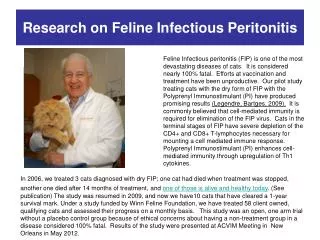
Feline Infectious Peritonitis FIP. Accession: 11114 and 13858. Skeeter 1.5 yo DSH with central vestibular signs. Thor 14 yo DSH; history of bicavitary effusion and heart murmur. FIP-Review. 1/200 new feline cases to American VTH

E N D
Feline Infectious PeritonitisFIP Accession: 11114 and 13858
Thor14 yo DSH; history of bicavitary effusion and heart murmur
FIP-Review • 1/200 new feline cases to American VTH • FIP is a fatal immune-mediated dz triggered by infection with a mutated feline coronavirus • 5% of FCoV infected cats develop FIP in a cattery situation, although infection rates are 50-90% • Non-mutated FCoV replicates in enterocytes and may cause diarrhea or transient resp signs • Single virulent mutation allows virus to replicate in macrophages
FIP-Review • Over half of the cats with FIP are younger than 12 months of age • Transmission of non-mutated FCoV: • Oronasal infection • Most commonly transmitted through virus-containing feces • Rarely transmitted through saliva (salivary shedding early in infection), by mutual grooming, by sharing the same food bowl, or through close contact. • Sneezed droplet transmission possible • Transplacental transmission can occur, but is rare
FIP-Review • Mutated FIP-causing FCoV has not been found in secretions or excretions of cats with FIP • Transmission of the mutated FIP causing FCoV not likely naturally, can be iatrogenic
Pathophysiology • Viral mutation leads to changes in the surface structures of the virus that allow the virus phagocytized by macrophages to bind to the ribosomes in these monocytes/macrophages and replicate there • Clinical signs manifest 2 wks to 2 years after mutation
Proposed mechanisms of pathogenesis • FCoV-infected macrophages leave the bloodstream and enable virus to enter the tissues, where attracts antibodies and fixes complement. Secondary inflammatory cell recruitment result in granuloma formation • Immune complexes exit circulation into vascular walls, fix complement, granuloma formation
Pathogenesis • Complement fixation • Release of vasoactive amines • Endothelial cell retraction • Increased vascular permeability w/ exudation of plasma proteins and development of modified transudate effusions
Forms of FIP • Effusive, exudative, wet form • fibrinous peritonitis, pleuritis, or pericarditis • 60% of cats with ascites have FIP • 14% of cats with pericardial effusion have FIP • Noneffusive, nonexudative, dry form • granulomatous changes in visceral organs, eye, and CNS • Mixed form • Differentiating forms has no prognostic significance, one form can progress to the other form
Sites of disease • CNS: ataxia, nystagmus, seizures most common • 75% have hydrocephalus • Ocular: Retinal cuffing, uveitis • Abdomen: Mesenteric LN, visceral organs or just bowel wall, esp ICJ
Diagnosis • CBC-incr/decr WBC; anemia (AIHA, Heinz bodies); decr platelets (DIC) • Chemistry-reflect organ involvement • Hyperglobulinemia-if serum A/G ratio<0.8 has 92% PPV for FIP • Cavity fluid-straw-colored modified transudate
Splenic US findings • 2 cases: Pyogranulomatoussplenitis secondary to FIP (2001) • Abdominal effusion • Large spleen, normal echogenicity • Irregular contour in one cat • Compared to dogs, cats have little periarterial lymphatic tissue, with the lymphatic tissue confined to nodules of white pulp.
MRI findings in Neuro FIP • Pachymeningeal enhancement, especially of brainstem and spinal cord • Hydrocephalus-obstructive • Marked ependymal enhancement • Periventricular contrast enhancement • The 4th ventricle was the most consistently affected (4/4), and the lateral ventricles were the least affected (1/4).
Clinical features of neurologic FIP • Histopathologic lesions in the brain consisted of meningitis, ependymitis or periventriculitis, and choroiditis of varying severity • Lesions most severe at the base of the cerebellum and brain stem, including the medulla oblongata.
Diagnosis? • Antibody tests-cannot differentiate enteric form and mutated form! • PCR tests cannot differentiate two forms! • Possibly helpful on CSF or effusion? • Not adeq studied, may be very helpful
Diagnosis • IFA-FIP antigen-effusion or tissue • 100% PPV on effusion, 57% NPV • “… intracellular FCoV antigen by immunofluorescence or immunohistochemistry is the only way to diagnose FIP definitively. This tool should be used whenever possible.” (Hartmann, 2005) • Histopathology/Necropsy • Pathognomonic lesions: localized perivascular mixed inflammation w/macrophages, neutrophils, lymphocytes, and plasma cells
References • Foley JE, Lapointe J, Koblik PD, Poland A, Pedersen NC. Diagnostic features of clinical neurologic feline infectious peritonitis. J Vet Intern Med 1998;415-423. • Hanson, JA, et al. Ultrasonographic appearance of splenic disease in 101 cats. Veterinary Radiology & Ultrasound, Vol. 42, No. 5, 2001, pp 441-445. • Hartmann K. Feline Infectious Peritonitis. Vet Clin Small Anim, 35, 2005. pp 39–79. • Mellema, JM et al. Meningeal enhancement on magnetic resonance imaging in 15 dogs and 3 cats. Veterinary Radiology & Ultrasound, Vol. 43, No. I , 2002. pp 10-15.