Download
1 / 153
1.55k likes | 1.88k Views
Acute Gastrointestinal Hemorrhage. Sirikan Yamada, M.D., F.R.C.S.T Assistant Professor Department of Surgery Faculty of Medicine, CMU Chiang Mai, Thailand. “Learning without thinking is useless. Thinking without learning is dangerous.”. - Confucius. Acute Gastrointestinal Hemorrhage.

E N D
Acute Gastrointestinal Hemorrhage Sirikan Yamada, M.D., F.R.C.S.T Assistant Professor Department of Surgery Faculty of Medicine, CMU Chiang Mai, Thailand
“Learning without thinking is useless.Thinking without learning is dangerous.” - Confucius
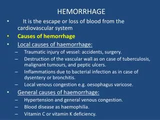
Acute Gastrointestinal Hemorrhage • Definition and Terminology • I> Upper gastrointestinal hemorrhage • (UGIH) • : Bleeding upon the ligament of treitz • Hematemesis : vomiting for fresh blood • shown active/ massive bleeding
Acute Gastrointestinal Hemorrhage • Definition and Terminology (cont) • Coffee ground: blood+gastric secretion • shown resent subside UGIH • Melena: Hb+acid= acid hematin, since 50cc • of blood 1000cc of blood caused melena persist For 5-7 days, and occult blood can be positive for 21 days
Acute Gastrointestinal Hemorrhage • II> Lower gastrointestinal hemorrhage • (LGIH): bleeding below ligament of Treitz • Hematochezia: means fresh blood, clot, or current jelly stool
Divisions of the stomach (From Zuidema G: Shackelford’s Surgery of the Alimentary Tract, 4th ed. Philadelphia, WB Saunders, 1995.)
Upper gastrointestinal hemorrhage(UGIH) • *** Guideline for approach and management non-variceal bleeding • - Related Surgical Anatomy and pathophysiology of Stomach and Duodenum • - Group of diseases caused UGIH and specific consideration • - Endoscopic and surgical management for UGIH
Upper gastrointestinal hemorrhage(UGIH) variceal bleeding • - Cirrhosis and portal hypertension • - Endoscopic diagnosis and management • - Surgical management
Lower gastrointestinal hemorrhage( LGIH) • - Relate Surgical Anatomy of small and large intestine • - Guideline for approach and treatment • - Group of diseases cause LGIH and specific consideration • - Historical background of investigation for localization and surgical management for LGIH
Why we have to learn ….. • Over all mortality rate is still high in upper GI hemorrhage, about 5-8% Dudnick R, Martin P, Friedman LS; management of bleeding ulcer. Med Clin North Am 75:948,1991
Blood supply to the stomach and duodenum with anatomical relationships to the spleen and pancreas. The stomach is reflected cephalad. (From Zuidema G: Shackelford’s Surgery of the Alimentary Tract, 4th ed. Philadelphia, WB Saunders, 1995.)
GASTRIC GLAND
Risk Factor for Peptic Ulcer Hemorrhage • Aspirin (ASA) : since 1899 • Non-steroidal antiinflammatory drug ( N-SAID) : has both Cyclooxygenase-1 and 2: COX - 1(house keeping enzyme) & COX 2( Co-enzyme) • Selective COX- 2 inhibitor: 1999 EX: Clelcoxib, Rofecoxib
COX theory • ASA – inhibit COX- 1, decrease Thromboxane& decrease prostaglandin caused of lost of protection for gastric mucosa, and decrease hemostasis • N-SAID- inhibits both COX-1 and COX-2 :results like in ASA user. Increase risk of complication in PU patients =6.1 (relative risk) and in recent GI bleeding patients= 13.5 (relative risk) • Selective COX-2 inhibitor: results more protection for gastric mucosal barrier, and hemostasis
Acute Gastrointestinal Hemorrhage ** Initial evaluation and treatment of patients with acute gastrointestinal hemorrhage
UPPER GI HEMORRHAGE • Initial Assessment • Initial Resuscitation • Critical care and monitoring • Definite diagnosis and management How should surgeons deal with and step up this complicatedproblem? Evidence based critical care, Paul Ellis Marick , 2001
A: General assessmentand Scoring to categorize the patients - Active or ongoing/ massive/ continue/ or intermittent bleeding B:Hx and PE (Cirrhotic patient or Non cirrhotic patient ) C:NG tube should be insertedto confirm the level of bleeding I. Initial Assessment
A. General Assessment • Hemodynamic assessment: BP, pulse, postural changes, peripheral perfusion • The presence of co morbid diseases • Estimation of blood loss by nasogastric tube intubation and hemodynamic response to fluid challenge ** remarked that - to use 2 L of crystalloid to stabilize v/s , blood loss is about 15-30% - If BP raises but fall again, blood loss is about 30-40% - If BP continues to fall, blood loss is more than 40%
Category of Hypovolemic Shock • Class I:Impending (< 10% of blood volumn loss) no symptom,pulse > 90-100 , BP normal • Class II: mild (10-20% of blood volumn loss) fainting, pallor, cool skin, BP drop, pulse>120 • Class III:modurate(20-30% of blood volumn loss): urine output -oliguria • Class IV: severe ( >40% of blood volumn loss) may caused unconcious and cardiac arrest
Category and scoring of patients • To evaluate and predict further ulcer hemorrhage • To select the method of management “ It is dictated by the rate of bleeding”
Clinical bleeding 1.Trace heme-positive stools and without severe anemia ( OPD) of cases 2.Visible blood, coffee ground, melena ( IPD/ further evaluation) 1+ 2 = 80 % of cases Fleischer D, et al Gastroenterology, 1983 3.Persistent or massive bleeding / referred due to rebleeding with hemodynamic instability (ICU) ** Massive/ ongoing bleeding is defined as loss of > 30% of estimated blood volume or bleeding required blood transfusion > 6 U/ 24 hours
Scoring to categorize the patients • Forrest classification severe, moderate, mild Lancet 1974 • Rockall Risk Scoring Gut 1996 • New Scoring System by Blatchford Lancet 2000 • Modified Rockall Score for both Non-variceal and Variceal bleeding AJG 2002
Rockall Scoring • Age • Shock • Co morbid disease ( cancer diseases) • Endoscopic diagnosis • Stigmata of recent hemorrhage Pre-endoscope score 0-7 Post –op endoscope score 0-11 * this scoring system is good to predict for the mortality rate much than rebleeding 0-3 : mortality rate = 0 – 11% 4-7 : mortality rate = 24- 27% • > 8 : motality rate = > 40% Rockall TA et al GUT 1996; 38: 316-21
New Scoring System by Blatchford • Admission Hb • BUN • Pulse • Systolic BP • Fainting or melena as chief complaint • Liver disease or cardiac disease • to predict the need for clinical interventions • But it is in only one study
High Risk ~Criteria • Host Factors • Age >60yr • Co-morbid conditions e.g. renal failure, cirrhosis, cardiovascular disease, COPD • Hemodynamic instability; mod to severe shock • Coagulopathy include drug-related • Bleeding character ; Active continue red blood from NG after irriagtion and red blood per rectum • Patient course; massive blood transfution> 4-6 units to maintain Hb in 24 hr , re-bleeding in 72 hr , return to have hemodynamic instability 2004 Concensus for Clinical Practice Guideline for the Management of Upper GI bleeding; สมาคมโรคทางเดินอาหารแห่งประเทศไทย
A: General assessment and Scoring to categorize the patients - Active or ongoing/ massive/ continue/ or intermittent bleeding B: Hx and PE(Cirrhotic patient or Non cirrhotic patient ) C: NG tube should be insertedto confirm the level of bleeding I. Initial Assessment
B. Take Hx and PE (Cirrhotic patient or Non cirrhotic patient ) - History taking of previous medication and underlying diseases/ anticoagulant usage. • Esophageal varices is more suspicious for 60% - 80% in severe upper GI bleeding with history of advanced liver disease or a history of previous variceal bleeding.
Prediction of UGI bleeding etiology Incidence(%) • Duodenal ulcer 24.3 • Gastric erosions 23.4 • Gastric ulcer 21.3 • Esophageal varices 10.3----------20% (in cirrhosis) • Malorry-Weiss tear 7.2 • Esophagitis 6.3 • Duodenitis 5.8 • Neoplasm 2.9 • Marginal( stomal) ulcer 1.8 • Esophageal ulcer 1.7 • Miscellaneous 6.8 Silverstein FE, Gilbert DA, Tadeseo FJ, et al, The national ASGE Survey on upper gastrointestinal bleeding Gastrointestinal Endoscopy, 1981
A: General assessment and Scoring to categorize the patients - Active or ongoing/ massive/ continue/ or intermittent bleeding B: Hx and PE (Cirrhotic patient or Non cirrhotic patient ) C: NG tubeshould be insertedto confirm the level of bleeding I. Initial Assessment
C. NG tube placement • Should perform in all UGI hemorrhage • to confirmation that it is the upper GI bleeding , monitoring of bleeding and , decompressed the stomach • No report that it may potentiate bleeding in case of esophageal varices, just careful in patients who had severe coagulopathy.
UGI LGI • Melena • Hematemasis or coffee ground • Maroon stool * • Red stool ** * • Guaiac test ( can positive more 2 weeks after bleeding stopped) * Bile was seen via NG tube ** Massive bleeding
NecessaryLaboratories • CBC,plt • BS, BUN, Cr, electrolyte • PT, PTT, bleeding time • LFT • G/M • EKG • CxR
II. Initial Resuscitation • How to do for good resuscitation? • When will we give blood transfusion ? • Which medication will be used?
Large- bore intravenous lines or central lines • NG tube aspiration (by hand) to decompress clot in stomach • Volume expansion with colloid or crystalloid • Transfusionof blood immediately if patient has hemodynamically unstable * Blood products are the most efficient volume expanders ** It take about 72 hours for Hct to reach its nadir; therefore, a normal or moderate low Hct does not exclude significant bleeding *** Conversely, minimally falling of Hct also representfluid disequilibrium much rather than continued bleeding
If patients have coagulopathy, they should be corrected. - PTT prolong > 1.5 times - Platelet < 50,000/ mm3 - FFP should be given after 6 unit of PRC and plt should add after 10 unit of PRC • Monitoring V/S, urine out put /hour • Airway protection in those who have alteration of consciousness or endotracheal intubations may facilitate to investigate and give treatment in these patients
Recommendation for empiric Acid- suppression therapy Traditionally treated, even before the cause is determined, with acid suppression therapy. Medications are extremely safe, although the efficacy of this practice has not been proven conclusively. Kupfer, et al Gastroenterol Clin of North Amer, 2000 I.V. Proton pump inhibitor is more effective than I.V. H 2 blocker in increasing intragastric pH Vasopressin should not be used due to its systemic side effect