Download
1 / 34
430 likes | 759 Views
Stable Angina, Guidelines & RACPC. Promoting Assessment & Treatment which is Structured, Systematic, Objective Evidence-based Appropriate In keeping with the patient’s wishes Risk stratification At presentation (“pre-test”) After non-invasive assessment (“post-test”)

E N D
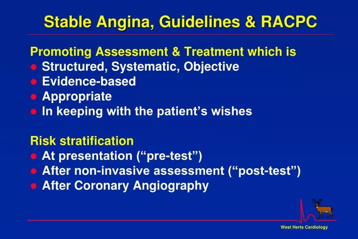
Stable Angina, Guidelines & RACPC Promoting Assessment & Treatment which is • Structured, Systematic, Objective • Evidence-based • Appropriate • In keeping with the patient’s wishes Risk stratification • At presentation (“pre-test”) • After non-invasive assessment (“post-test”) • After Coronary Angiography
Angina : NSF Standards Standard 8 People with symptoms of angina or suspected anginashould receive appropriate investigation and treatment to relieve their pain and reduce their risk of coronary events. Standard 9 People with angina that is increasing in frequency or severity should be referred to a cardiologist urgently or, for those at greatest risk, as an emergency. Standard 10 NHS Trusts shouldput in place hospital-wide systems of care so that patients with suspected or confirmed coronary heart disease receive timely and appropriate investigation and treatment to relieve their symptoms and reduce their risk of subsequent coronary events.
Arterial blood flow Arterial wall • Endothelium • Intima • Muscular wall Blood • Red cells • White cells • Platelets • Plasma(with O2 & Nutrients) • Clotting factors • Cholesterol • Toxins.. Alt-F4 to close movie Space/ controls playback
Endothelial Dysfunction Atherosclerosis Timeline Foam Cells Fatty Streak Intermediate Lesion Atheroma Fibrous Plaque Complicated Lesion / Rupture From First Decade From Third Decade From Fourth Decade Adapted from Pepine CJ. Am J Cardiol. 1998;82(suppl 104).
Clinical Manifestations of Atherothrombosis Transient ischemic attack (TIA) Ischaemic stroke (CVA) Acute Coronary Syndrome (ACS) Myocardial infarction (MI) Angina AAA Renovascular Disease Peripheral Vascular Disease (PVD)
Angina: Prognosis 100 90 Webster: 1960-5 Severe CHD Kannel : 1949-66 Framingham Men, >50yrs, Angina Podrid : 1981 ExECG +, Mild Angina Podrid 80 % Survival 70 Kannel Webster 60 2 4 6 8 10 Years of Follow-up Goldman L et al Am J Cardiol 1983;51:449-52
Progression of Vascular Disease Asymptomatic, or Stable Vascular Disease egStable Angina Stable AtheromatousPlaque Unstable Plaque Complications • Sudden Death • ACS • Myocardial Infarction • Heart Failure • Stroke • etc, etc Risk Factors Genetics
If demand for blood supply cannot be met, muscle becomes ischaemic Stable Angina in CHD Narrowing of Coronary artery limits blood supply to heart muscle
Stable Angina: Symptoms "There is a disorder of the breast with strong and peculiar symptoms considerable for the kind of danger belonging to it. The seat of it and the sense of strangling and anxiety with which it is attended may make it not improperly called angina pectoris. They who are afflicted with it are seized while they are walking with a painful and most disagreeable sensation in the breast ... But the moment they stand still all this uneasiness vanishes. If the pain continues, patients suddenly fall down and perish almost immediately.” William Heberden London Medical Transactions 1772
Effects of Myocardial Ischaemia Myocardial Ischaemia TransientLV Dysfunction ProgressiveLV dysfunction Angina Breathlessness Arrhythmia Most Myocardial ischaemia is painless (“Silent”) …. Sudden Death
Coronary AtheroThrombosis gradual, progressive sudden, ± occlusive Other causes of Coronary flow active spasm lack of vasodilatation Cold Anaemia Carbon Monoxide High Altitude Increased Heart Rate Exercise, stress Smoking Increased LV stress LVH, Hypertension Aortic Stenosis, HCM Cold Food Hyperthyroidism Causes of Myocardial Ischaemia Reduced Oxygen Supply Increased Oxygen Demand • : Effects of cigarette smoking
Evaluation and Diagnosis • In patients presenting with chest pain • detailed symptom history • focused physical examination • directed risk-factor assessment • Estimate the probability of significant CHD • ifintermediate or high: refer to RACPC • Objective assessment (egExECG) is for: • Diagnosis of myocardial ischaemia • Assessment of severity & pathophysiology • Assessment of prognosis
Rapid Access Chest Pain Clinics “One-stop” assessment of stable patients Recent (<6 months) onset of exertional chest pain, intermediate-high risk of angina Known CHD which was stable (eg after PTCA or CABG) now symptomatic again < 2 week wait to clinic
Classification of Chest Pain Estimating the Probability of CHD from History of Chest Pain • Precipitated by exercise • Brief duration (<15 minutes) • Relieved promptly by rest or GTN • Central chest location • Radiates to Jaw, Throat, or L Arm • Absence of other causes for pain CHD If only ONE criterion + = Non Anginal pain : < 30% If only criteria 4-6 +,or any TWO + = Chest Pain ? Cause : 30-70% If only criteria 1-3 +,or any FOUR + = Typical Angina : > 70% Diamond GA, Forester JS. NEJM 1979;300:1350-8 Patterson RE, et al JACC 1989;13:1653-65
Pre-test probability of CHD: Duke Score • By combining • Classification of Chest Pain • CHD risk factors (including ECG) • a more accurate prediction of the (pre-test) probability of significant CHD can be generated • Structured, systematic, objective assessment • Easy to use on web, PC or PDA Pryor DB et al Ann Int Med 1993;118:81-90
Pre-test probability of CHD: Duke Score Probability of CHD 50% Multiple risk factors Probability of CHD increased to 88% Pryor DB et al Ann Int Med 1993;118:81-90
Pre-test probability of CHD: Duke Score Probability of CHD 50% Few risk factors Probability of CHD decreased to 15% Pryor DB et al Ann Int Med 1993;118:81-90
Pre-test probability of CHD: Duke Score Demonstration of • web-based RACPC referral form • automatic risk assessment www.westhertshospitals.nhs.uk/whc Risk calculators → RACPC Referral
Rapid Access Chest Pain Clinics Not every patient with chest pain is suitable Acute MI / Unstable Angina: CCU Stable Angina with mod-high prob of CHD Chest Pain ? Cause with mod prob of CHD Atypical Pain with low prob of CHD: ?? OP ACS / MI, Heart Failure, Valve Disease, Palpitations Anaemia, AF, Digoxin, LVH++, LBBB, can’t walk
Evidence-based Management of Angina • Careful assessment • ? Underlying cause • ? Risk factors : Smoking, Lipids, BP, DM • ? Prognosis : Exercise ECG • Treatment • Stop smoking, lose weight, healthy diet • Aspirin, + Statin and ACEI as appropriate for 2y prevention • Blocker if possible (else Verapamil or Diltiazem) • Nicorandil or Nitrate, using GTN prophylactically • Consider Angiography Revascularisation
Stable Angina Guidelines Gibbons et al JACC 1999;7:2092–197
Prognostic Markers in Exercise Testing The Duke Treadmill Score= • exercise time in minutes on Bruce Protocol • minus 5 x the ST-segment deviation during or after exercise (mm) • 4 x the angina index 0 if there is no angina 1 if angina occurs, and 2 if angina is the reason for stopping the test • works well for both inpatients and outpatients, and equally well for men and women Mark DB et al NEJM 1991;325:849-53Shaw LJ et al Circulation 1998;98:1622-30
Duke Treadmill Score Survival According to Risk Groups 4 -Year Annual Risk Group (Score) Total Survival Mortality Low ( +5) 62% 99% 0.25% Moderate (-10 to +4) 34% 95% 1.25% High (< -10) 4% 79% 5.00% Mark DB et al NEJM 1991;325:849-53Shaw LJ et al Circulation 1998;98:1622-30
Use of Duke Treadmill Score Predicted average RecommendedRisk score annual mortality treatment low <1% per year Medical therapy intermediate 1% to 3% Cardiac Catheterization ? Stress imaging high-risk >3% per year Cardiac Catheterization * <5% pt with low-risk treadmill score will be identified as high risk after imaging* those with known LV dysfunction should have cardiac catheterization Mark DB et al NEJM 1991;325:849-53Shaw LJ et al Circulation 1998;98:1622-30
Coronary Revascularisation: 1 Limiting Angina despite Medical treatment Recent MIor Unstable Angina Non-Invasive assessment (eg ExECG) indicates Risk High Risk Angiography Low Risk Medical treatment
Coronary Revascularisation: 2 Limiting Angina despite Medical treatment Recent MIor Unstable Angina Non-Invasive assessment (eg ExECG) indicates Risk High Risk Angiography Normal or Mild CHD 1-2 vessel CHD 3 vessel CHD or LMS Low Risk Medical treatment PTCA ± Stent MICAS CABG Balance Risk v Benefit
Risk Stratification With Coronary Angiography • Extent and severity of coronary disease and LV dysfunction are the most powerful clinical predictors of long-term outcome • proximal coronary stenoses • severe left main coronary artery stenosis • In the CASS registry of medically treated patients, the 12-year survival rate by Coronary arteriesEjection fraction normal coronary arteries 91% 50% to 100% 73%one-vessel disease 74% 35% to 49% 54%two-vessel disease 59% <35% 21%three-vessel disease 40% CASS Circulation 1994;90:2645-57
Prognosis of CHD by severity at Angio 100 80 Distal coronary disease 1 vessel CHD 2 vessel CHD 3 vessel CHD Left Mainstem Stenosis 60 Probability of Survival (%) 40 20 0 1 2 3 4 5 Years Balcon R, Davies S The management of stable angina RCP;1994:p61
Canadian Cardiovascular Society Classification of Stable Angina severity Class I: Ordinary physical activity does not cause anginaNo angina on ordinary walking or climbing stairs.Angina with strenuous or rapid or prolonged exertion at work or recreation. Class II: Slight limitation of ordinary activityAngina on walking or climbing stairs rapidly, walking uphill, walking or stair climbing after meals, in cold, or in wind, or when under emotional stress, or only during the few hours after awakening; walking more than 100-200m on the level or climbing more than one flight of stairs at a normal pace and in normal conditions. Class III: Marked limitation of ordinary physical activityAngina on walking 100-200m on the level or climbing one flight of stairs in normal conditions and at normal pace. Class IV: Inability to carry on any physical activity without discomfort Anginal syndrome may be present at rest. Campeau L. Circulation 1976;54:522–523