Download

1 / 66
660 likes | 666 Views
Learn about the female reproductive system, functions of the uterus, characteristics of sperm and ovum, the process of ovulation, and the three stages of prenatal development.

E N D
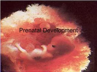
FEM 3101Developmental Psychology: Children and AdolescentsPJJ - FACE TO FACE 2SEMESTER 2SESSION 2012-201324 FEBRUARY 2013 SITINOR/FEM3101/FEBRUARI 2013/PJJ
PRENATAL DEVELOPMENT • Reproductive systems • Stages in prenatal development • Context of development • Influences on prenatal development SITINOR/FEM3101/FEBRUARI 2013/PJJ
The Female Reproductive System • Uterus • A muscular chamber about the size and shape of a pear. • Located in a woman's abdomen, is a hollow, elastic reproductive organ, where a baby develops during pregnancy. SITINOR/FEM3101/FEBRUARI 2013/PJJ
Female anatomy • The uterus - is a major female hormone-responsive reproductive sex organ • Within the uterus fetus develops during gestation. • The term uterus =womb. • One end, the cervix, opens into the vagina; the other is connected on both sides to the Fallopian tubes. SITINOR/FEM3101/FEBRUARI 2013/PJJ
Sperm • Sperm is the male reproductive cell Chief Characteristics: • Tightly packed tip (acrosome) that contains 23 chromosomes that carry genetic information • Short neck region • Trail to propel it in its search for the ovum • Microscopic Fact: • Remains capable of fertilizing egg for 24-48 hours after ejaculation • Of 200 million sperm that enter the vagina, only about 200 survive the journey to the fallopian tubes, where fertilization occurs • Males, at birth, have in their testes those cells that will eventually produce sperm SITINOR/FEM3101/FEBRUARI 2013/PJJ
Ovum (Egg) • The ovum is the female reproductive cell Chief Characteristics: • Round • .01 mm in diameter • Consistency of stiff jelly • Contributes 23 chromosomes Fact: • Females already have 1-2 million primal eggs at birth • Eggs usually fertilized about 12 hours after discharged from the ovary or they die within 12-24 hours SITINOR/FEM3101/FEBRUARI 2013/PJJ
ovulation • When a young woman reaches puberty, she begins to ovulate • a process in which a mature egg cell (also called an ovum), ready for fertilization by a sperm cell, is released from one of the ovaries SITINOR/FEM3101/FEBRUARI 2013/PJJ
Her body prepares for a potential pregnancy every cycle, whether or not she want to actually conceive. • Under the influence of Follicle Stimulating Hormone (FSH), about 15 to 20 eggs start to mature in each ovary. • Although it averages about two weeks, the process to release an egg can take anywhere from about eight days to a month or longer to complete. SITINOR/FEM3101/FEBRUARI 2013/PJJ
menstrual cycle • Ovulation occurs 14 days before the next menstruation. • As the average menstrual cycle lasts 28 days (starting with the first day of one period and ending with the first day of the next menstrual period), most women ovulate on day 14. SITINOR/FEM3101/FEBRUARI 2013/PJJ
A menstrual cycle can vary between 21 to 38 days. • A woman is generally most fertile (able to become pregnant) a few days before, during, and after ovulation. • The corpus luteum remains behind on the interior ovarian wall, and starts releasing progesterone. • Progesterone quickly stops the release of all other eggs until the next cycle. The corpus luteum has a finite lifespan, of about 12 to 16 days. SITINOR/FEM3101/FEBRUARI 2013/PJJ
Menstruation • If the egg does not become fertilized as it travels down the fallopian tube on its way to the uterus, the endometrium (lining of the uterus) is shed and passes through the vagina (the passageway through which fluid passes out of the body during menstrual periods; also called the birth canal), a process called menstruation. SITINOR/FEM3101/FEBRUARI 2013/PJJ
Pregnancy • If the egg is fertilized by a sperm cell as it travels down the fallopian tube, then pregnancy occurs, it becomes attached to the lining of the uterus SITINOR/FEM3101/FEBRUARI 2013/PJJ
In order for conception to occur, though, there must be three factors present: • the egg, • the sperm • a medium in which the sperm can travel to reach the fallopian tubes. • Women produce cervical fluid under the influence of increasing levels of estrogen in the first part of the cycle. • Sperms can live up to five days in fertile quality cervical fluid SITINOR/FEM3101/FEBRUARI 2013/PJJ
3 Stages in prenatal development: • Germinal stage • Embryonic stage • Fetal stage SITINOR/FEM3101/FEBRUARI 2013/PJJ
Conception: First phase of development – Period of the zygote • The development of a single human being begins with conception when a single sperm cell from the male unites with an egg from a female and forms a single cell called a zygote. • Once conception has occurred, the ovum continues down the fallopian tube. Then, it implants itself in the wall of its uterus. • This is the first phase of development and it is known as the period of the zygote. SITINOR/FEM3101/FEBRUARI 2013/PJJ
Business Driven • Technology Oriented • Sustainable Development • Environmental Friendly Conception The period of the zygote (Fertilization to 2 weeks) • This period lasts about 2 weeks. • The term zygote is used to refer to the organism throughout this period. • In the early stages, the mass of cell is undifferentiated. • However, about four days after conception some differentiation begins, at which point the organism is called blastocyst. SITINOR/FEM3101/FEBRUARI 2013/PJJ
Conception The period of the zygote (Fertilization to 2 weeks) • A blastocyst is a hollow ball of cells that has developed from the fertilized egg. • During this time, cells begin to differentiate. • By the end of the period of the zygote, the developing organisms has found food and shelter in the uterus and developed into the embryonic stage. SITINOR/FEM3101/FEBRUARI 2013/PJJ
Conception The embryonic stage (2 to 8 weeks) • The second major phase of prenatal development (the embryo) begins with completion of implantation • It continues for another six weeks until the various support structures are fully formed and all the major organ systems have been laid down in at least rudimentary form. SITINOR/FEM3101/FEBRUARI 2013/PJJ
Conception The embryonic stage (2 to 8 weeks) • The embryo is especially vulnerable to interference with healthy development. • This stage begins at week 3 and ends in the second month (week 8) of conception. SITINOR/FEM3101/FEBRUARI 2013/PJJ
Conception The embryonic stage (2 to 8 weeks) • The embryo’s circulatory is connected to the placenta through the umbilical cord. • The placenta is connected to both the mother’s and the embryo’s (fetus’s) blood system, but the two systems are not directly connected. • Small molecules pass back and forth through this large filtering system, but large ones cannot. SITINOR/FEM3101/FEBRUARI 2013/PJJ
Conception The embryonic stage (2 to 8 weeks) • So nutrients such as oxygen, proteins, sugars, and vitamins from the maternal blood pass through to the embryo or fetus, while digestive wastes and carbon dioxide from the infant’s blood pass back through to the mother, whose own body can eliminate them. • The period from the ninth week of conception until the end of pregnancy is called the fetal stage or the period of the fetus. The embryo is called fetus when the first bone cell appears. SITINOR/FEM3101/FEBRUARI 2013/PJJ
Conception • Business Driven • Technology Oriented • Sustainable Development • Environmental Friendly The period of the fetus (8 weeks to birth) • This is the longest prenatal period. • The seven months of the fetal stage involve primarily a process of refining all the primitive organ systems already in place. • At the end of the embryonic period, the main parts exist in some basic form; the next seven month are for the finishing process. SITINOR/FEM3101/FEBRUARI 2013/PJJ
Conception • Business Driven • Technology Oriented • Sustainable Development • Environmental Friendly The period of the fetus (8 weeks to birth) • During this phase, the organisms begins to increase rapidly in size, about 20 times its previous length; organs and body systems become more complex. • This period is divided into second trimester and third trimester. • Table 2.1 displays milestones of prenatal development. • Figure 2.1 shows the growth of the brain during the prenatal period. SITINOR/FEM3101/FEBRUARI 2013/PJJ
Table 2.1 Milestones of Prenatal Development • Business Driven • Technology Oriented • Sustainable Development • Environmental Friendly SITINOR/FEM3101/FEBRUARI 2013/PJJ
Table 2.1 Milestones of Prenatal Development • Business Driven • Technology Oriented • Sustainable Development • Environmental Friendly SITINOR/FEM3101/FEBRUARI 2013/PJJ
Table 2.1 Milestones of Prenatal Development • Business Driven • Technology Oriented • Sustainable Development • Environmental Friendly SITINOR/FEM3101/FEBRUARI 2013/PJJ
Table 2.1 Milestones of Prenatal Development • Business Driven • Technology Oriented • Sustainable Development • Environmental Friendly SITINOR/FEM3101/FEBRUARI 2013/PJJ
Fertilization • Prenatal development begins when the ovum and sperm unite (i.e., fertilization), creating a new and separate cell called the Zygote SITINOR/FEM3101/FEBRUARI 2013/PJJ
FIRST CELL DIVISION Immediately the cell begins to duplicate, taking approximately 30 hours to complete the first cell division. SITINOR/FEM3101/FEBRUARI 2013/PJJ
At an increasingly faster rate, new cells are added until they form a hollow, fluid-filled ball, called a blastocyst (about 4 or five days after conception). Approximately 60 to 70 cells form the blastocyst. Those on the inside (called the embryonic disk) will become the new organism whereas those on the outside will provide the protective covering. BLASTOCYST SITINOR/FEM3101/FEBRUARI 2013/PJJ
Around the seventh or ninth day, the blastocyst implants itself into the uterine lining. The protective covering quickly develops into the amnion, surrounding the organism in amniotic fluid. A yolk sac also develops, producing blood cells until the liver, spleen, and bone marrow is mature. IMPLANTATION SITINOR/FEM3101/FEBRUARI 2013/PJJ
The Period of the embryo lasts from about 2 weeks until about the 8th week of pregnancy. During this time, the groundwork for all body structures and organs is laid. 8 WEEKS FROM CONCEPTION EMBRYO: 5 WEEKS SITINOR/FEM3101/FEBRUARI 2013/PJJ
Even before the mother knows she is pregnant: the heart has begun to pump blood; the muscles, backbone, and ribs have begun to appear; and tiny buds have developed into arms, legs, fingers, and toes. Embryo: 6 Weeks SITINOR/FEM3101/FEBRUARI 2013/PJJ
By the 7th week, the liver and spleen begins producing blood cells and the heart has developed separate chambers. At this time, the tiny organism shows sensitivity to touch and freely moves about in the amniotic sac. However, at less than an inch long and only an ounce in weight, the organism is still too tiny for any movements to be felt by the mother. EMBRYO: 7 WEEKS SITINOR/FEM3101/FEBRUARI 2013/PJJ
By the end of the embryonic period, the internal organs as well as external structures have become more distinct. Illustration: The development of the eyes. "The eyes form on stems that have grown from either side of the front of the brain out to the skin on the face At first, the eyes are mere indentations on the side of the head, but they develop rapidly through seven (top), eight (middle), and 10 (bottom) weeks of pregnancy. By three months, the eyelids form, and then close for a few months over the newly formed eyes." (text by Your Growing Child) 8 WEEKS 9 WEEKS 10 WEEKS SITINOR/FEM3101/FEBRUARI 2013/PJJ
The 3rd month of pregnancy marks the end of the first trimester for the mother, and the end of the first month of the Fetal Period. The fetal period is the longest prenatal period, lasting from the ninth week to the end of pregnancy. FETUS – 3 MONTHS • During the third month, the organs, muscles, and nervous system become connected and organized. • The fetus can kick, bend its arms, make a fist, open its mouth, and can even suck its thumb. • The skin of the fetus is thin and transparent. Thus, the internal organs and features can still easily be seen with an internal camera SITINOR/FEM3101/FEBRUARI 2013/PJJ
During the 4th month - vernix (a white, cheeselike substance) covers the entire body of the fetus. The vernix protects the skin from chapping during the several months that the fetus is in the amniotic fluid. A white, downy hair called lanugo also covers the fetus' body, which helps the vernix stick to the skin. The fetus has grown large enough that the movements can sometimes be felt by the mother. Often felt like a flutter or a "flip-flop“. These first movements that can be felt by the mother is called quickening. 11-14 weeks FETUS - FOUR MONTH SITINOR/FEM3101/FEBRUARI 2013/PJJ
At 22 weeks, the fetus weighs a little over 1 pound, and is about 1 foot in length. At this time, the movements can clearly be felt by the mother and by others who place their hands over the mother's abdomen. The fetus also shows a sensitivity to light and can be stimulated and irritated. However, it still has a long way to go before it is mature enough to survive outside of the womb. Although there are a few cases of infants being born and surviving at this time, the chance of survival (and without later complications), is very slim. FETUS – 5 MONTHS SITINOR/FEM3101/FEBRUARI 2013/PJJ
The 6th month marks the beginning of the third trimester for the mother. If born during this trimester, the fetus has a chance survival. The point in which it can first survive is referred to as the age of viability and occurs sometime between 22 and 26 weeks. FETUS – 6 MONTHS SITINOR/FEM3101/FEBRUARI 2013/PJJ
At only 3-4 pounds, the 7 month old fetus has yet another 3-4 pounds to go before reaching the average 7.5 pounds. During this time, the brain continues to develop at at increasingly fast rate. By 7 months, the fetus clearly responds to sounds outside of the womb, developing a preference for the tone and rythm of its mother's voice. FETUS – 7 MONTHS SITINOR/FEM3101/FEBRUARI 2013/PJJ
By the 8th month, the fetus has little room for large movements. During this month, a layer of fat is added that will assist with temperature regulation. The lungs however, still remain immature. If born at this time, the infant will likely require some help with breathing. It is not until the 9th month that the lungs are mature enough to regulate breathing without assistance. FETUS – 8 MONTHS SITINOR/FEM3101/FEBRUARI 2013/PJJ
A CHILD IS BORN One minute, and again at five minutes after birth, the infant is assessed using the APGAR scale. On average, the newborn infant weighs 7.5 pounds and is 20 inches long. SITINOR/FEM3101/FEBRUARI 2013/PJJ
A majority of the newborn's first month is spent sleeping, waking every few hours to be fed. BABY – 3 WEEKS OLD SITINOR/FEM3101/FEBRUARI 2013/PJJ
Embrio : blastosis burrows into the uterine lining SITINOR/FEM3101/FEBRUARI 2013/PJJ
As soon as the fertilized egg burrows into the lining, it starts releasing a pregnancy hormone, HCG (Human Chorionic Gonadotropin) which sends a message back to the corpus luteum left behind on the ovarian wall. • HCG signals the corpus luteum to remain alive beyond its usual maximum of 16 days and continuing to release progesterone long enough to sustain the nourishing lining. • After several months, the placenta takes over, not only maintaining the endometrium, but providing all the oxygen and nutrients the fetus needs to thrive. SITINOR/FEM3101/FEBRUARI 2013/PJJ
Cells Division • There are two type of cell division • Mitosis and meiosis • Reproductive cells divide through meiosis process, while all other body cells divide through the mitosis process SITINOR/FEM3101/FEBRUARI 2013/PJJ
Cells Division • Mitosis is cell division that results in the duplication of cells; the daughter cells genetic copies of the parent cell. This cell multiplication allows for replacement of old cells, tissue repair, growth and development. • Mitosis • The creation of new cells through duplication of chromosomes & divisions of cells cells duplicates (From 1 24 16 32, etc) • Cells developed into organs, brain, heart etc. • Growth & Development • You grew from a zygote, or fertilized egg (the fusion of two cells: an egg and a sperm) into an organism with trillions of specialized cells. • Mitosis is the process that enabled you to grow and develop after that fateful meeting of ovum and sperm became ‘you’. • Cell Replacement • Cells must divide in order for an organism to grow and develop, but cell division is also required for maintenance, cell turnover and replacement. SITINOR/FEM3101/FEBRUARI 2013/PJJ
Meiosis is Sex Cell (Gamete) Formation • In sexually reproducing organisms, some cells are able to divide by another method called meiosis. • Meiosis is a complex process by which gametes form; involves duplication and division of reproductive cells and their chromosomes. • The number of chromosomes in cells divide into two’s, and each set of cell will receive 1 from each sets of chromosomes makes up 23 sets. • This type of cell division results in the production of gametes (eggs or sperm). • Meiosis is much more complex than mitosis involves the duplication and subsequent division of chromosomes, meiosis involves two divisions of genetic material. As is the case in mitosis, in meiosis the cell duplicates its chromosome number prior to beginning cellular division. Then nuclear division, the sorting out of the genetic material, begins, and unfolds over the course of 2 cellular divisions that result in 4 gametes. SITINOR/FEM3101/FEBRUARI 2013/PJJ
Meiosis is Sex Cell (Gamete) Formation • Gametes & Gonads • Gametes are haploid (1n) with half the number of chromosomes than the progenitor cell that they arose from. These haploid sex cells arise in specialized reproductive tissue called the gonads. Ovaries (female gonads) and testes (male gonads) are the sites of meiosis. • Fertilization & Development • Sexual reproduction results in the merging of sperm and egg at fertilization, and brings the chromosome count back to the 2n diploid number necessary for a zygote to have complete genetic information; 2 sets of genetic instructions in 23 pairs of chromosomes. • As cells divide, the zygote develops and grows into an embryo, fetus and beyond. These 23 pairs of chromosomes are duplicated with every cell division, and are the genetic material inside nearly every cell of the body. SITINOR/FEM3101/FEBRUARI 2013/PJJ