Download
1 / 1
10 likes | 61 Views
Children’s Environmental Health Science Core Center. INTRODUCTION:

E N D
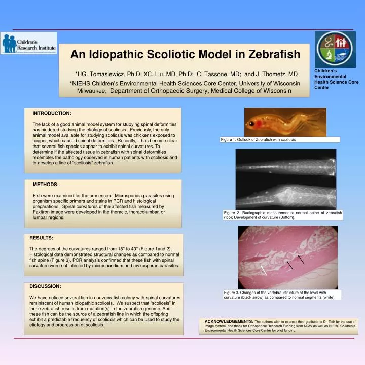
Children’s Environmental Health Science Core Center • INTRODUCTION: • The lack of a good animal model system for studying spinal deformities has hindered studying the etiology of scoliosis. Previously, the only animal model available for studying scoliosis was chickens exposed to copper, which caused spinal deformities. Recently, it has become clear that several fish species appear to exhibit spinal curvatures. To determine if the affected tissue in zebrafish with spinal deformities resembles the pathology observed in human patients with scoliosis and to develop a line of “scoliosis” zebrafish. An Idiopathic Scoliotic Model in Zebrafish *HG. Tomasiewicz, Ph.D; XC.Liu, MD, Ph.D; C.Tassone, MD; and J.Thometz, MD *NIEHS Children’s Environmental Health Sciences Core Center, University of Wisconsin Milwaukee; Department of Orthopaedic Surgery, Medical College of Wisconsin Figure 1. Outlook of Zebrafish with scoliosis. METHODS: Fish were examined for the presence of Microsporidia parasites using organism specific primers and stains in PCR and histological preparations. Spinal curvatures of the affected fish measured by Faxitron image were developed in the thoracic, thoracolumbar, or lumbar regions. Figure 2. Radiographic measurements: normal spine of zebrafish (top); Development of curvature (Bottom). RESULTS: The degrees of the curvatures ranged from 18° to 40° (Figure 1and 2). Histological data demonstrated structural changes as compared to normal fish spine (Figure 3). PCR analysis confirmed that these fish with spinal curvature were not infected by microsporidium and myxosporan parasites. DISCUSSION: We have noticed several fish in our zebrafish colony with spinal curvatures reminiscent of human idiopathic scoliosis. We suspect that “scoliosis” in these zebrafish results from mutation(s) in the zebrafish genome. And these fish can be the source of a zebrafish line in which the offspring exhibit a predictable frequency of scoliosis which can be used to study the etiology and progression of scoliosis. Figure 3. Changes of the vertebral structure at the level with curvature (black arrow) as compared to normal segments (white). ACKNOWLEDGEMENTS:The authors wish to express their gratitude to Dr. Toth for the use of image system, and thank for Orthopaedic Research Funding from MCW as well as NIEHS Children’s Environmental Health Sciences Core Center for pilot funding.