Download
1 / 1
10 likes | 139 Views
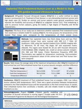
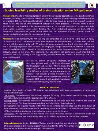
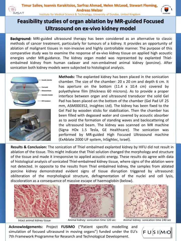
Timur Saliev , Ioannis Karakitsios , Sarfraz Ahmad, H elen McLeod, Stewart Fleming, Andreas Melzer Institute for Medical Science & Technology, University of Dundee, United Kingdom . Feasibility studies of organ ablation by MR-guided Focused Ultrasound on ex-vivo kidney model.

E N D
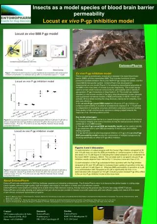
TimurSaliev, IoannisKarakitsios, Sarfraz Ahmad, Helen McLeod, Stewart Fleming,Andreas Melzer Institute for Medical Science & Technology, University of Dundee, United Kingdom Feasibility studies of organ ablation by MR-guided Focused Ultrasound on ex-vivo kidney model Background: MRI-guided ultrasound therapy has been considered as an alternative to classic methods of cancer treatment, particularly for tumours of a kidney. It provides an opportunity of ablation of malignant tissues in non-invasive and highly controllable manner. The purpose of this comparative study was to examine the response of ex-vivo kidney tissue on applied high acoustic energies under MR-guidance. The kidney organ model was represented by explanted Thiel-embalmed kidney from human cadaver and non-embalmed animal kidney (porcine). After sonication both kidney models were subjected to histological analysis. Methods: The explanted kidney has been placed in the sonication chamber. The size of the chamber: 20 x 20 cm and depth 6 cm. It has aperture on the bottom (11.4 x 10.4 cm) covered by polyethylene film (thickness 60 microns). As to provide a proper interface between organ and ultrasound transducer the solid Gel Pad has been placed on the bottom of the chamber (Gel Pad UF 25 mm, ASM000352, Insightec Ltd). The kidney has been fixed to the Gel Pad by wooden sticks for stabilization. Then the chamber has been filled with degassed water and covered by acoustic absorber as to avoid the formation of standing waves and backscattering of the ultrasound beam. The kidney was scanned on MR machine (SignaHDx 1.5 Tesla, GE Healthcare). The sonication was performed by MR-guided High Focused Ultrasound machine (ExAblate 2000 UF system, InSightec, Israel). Results & Conclusion: The sonication of Thiel embalmed explanted kidney by HIFU did not result in ablation of the tissue. This might indicate that Thiel solution changed the morphology and structure of the tissue and made it irresponsive to applied acoustic energy. These results do agree with data of histological analysis of sonicatedThiel-embalmed kidney tissue, where signs of the ablation were not detected. In opposite to the results from Thiel embalmed kidney, the samples from ablated porcine kidney demonstrated evident signs of tissue disruption triggered by ultrasound: obliteration of the morphological structure, defragmentation of the nuclei and cell lysis, discoloration as a consequence of massive escape of haemoglobin (below). Animal kidney: sonication time 120 sec Animal kidney: sonication time 240 sec Intact animal kidney tissue Acknowledgements: Project FUSIMO (“Patient specific modelling and simulation of focused ultrasound in moving organs”) funded under the EU's 7th Framework Programme for Research and Technological Development.