Download
1 / 28
300 likes | 604 Views
Male Anatomy. When you have completed this study you should be able to:. Recognize and identify the various parts of the male reproductive system Relate function to structure of each part Distinguish differences between species Recognize the internal structures of the male reproductive system

E N D
When you have completed this study you should be able to: • Recognize and identify the various parts of the male reproductive system • Relate function to structure of each part • Distinguish differences between species • Recognize the internal structures of the male reproductive system • Relate function to the internal structures
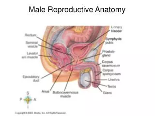
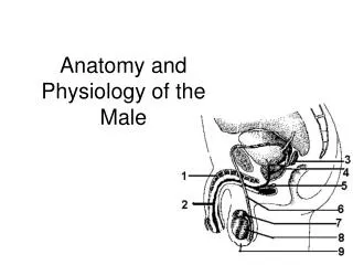
Anatomy of the Male Reproductive System • Functions to produce and deliver viable gametes to the female reproductive tract. • Scrotum • Testes • Epididymis • Spermatic cords • Accessory sex glands • Muscles • Penis
Scrotum • Two-lobed pouch that houses the testes • Aids in protection • Temperature regulation • Movement • Concentric layers Consisting of: • Skin • Tunica Dartos muscle • Fascia • Tunica Vaginalis
Testes • Male gonads • Function to produce male gametes • Spermatozoa • Produce male hormones (androgens) • Testosterone
Important for proper development of spermatozoa and for temperature control Abdominal cavity Gubernaculum Inguinal ring Scrotum Testicular Descent
Cryptorchidism Failure of one or both of the testes to descend into the scrotum (heritable trait) • Unilateral – When one testis does not descend into the scrotum. The testis that descends into the scrotum is fertile, however, reduced sperm concentrations usually result. • Bilateral – When neither testes descend into the scrotum. Results in sterility due to the elevated temperature.
Thermoregulation of the Testes Testes should be 4˚- 6˚C cooler than body temperature
Epididymis • Highly convoluted duct attached to the tunica albuginea that functions in: • Sperm transport • Spermatozoa maturation • Concentration of spermatozoa • Storage reservoir
Epididymis • Three segments • Caput • Contains efferent ducts • Fluid resorption • Corpus • Maturation • Cauda • Storage
Accessory Sex Glands • Ampullae • Vesicular Glands • Seminal vesicles • Prostate • Bulbourethral glands • Cowpers glands
Penis • Organ of copulation (3 parts) 1. Root (crus penis) 2. Body (corpus) 3. Glans penis
Penis • Corpus cavernosum • Majority of interior penile shaft • Spongy erectile tissue • Smooth muscle • Corpus spongiosum • Surrounds urethra • Extends to glans • Very prominent in stallion - belling
Muscles of the Penis • Retractor penis • Contracted/relaxed state • Ischiocavernosus • Inserts on crus penis • Important in erection • Compresses crus penis • Bulbospongiosus • Overlaps root of penis • Covers bulbourethral glands • Urethralis • Encloses pelvic urethra • Covers bulbourethral glands
Fibroelastic Penis Vs. Vascular Penis Fibroelastic: composed of a fiberous elastic type tissue that extends in length to create an erection rather than in diameter (as does the vascular type penis). Vascular: composed of corpus cavernosum (dorsal) and corpus spongiosum (ventral) erectile tissues. During an erection, the corpus cavernosum and the corpus spongiosum fill with blood causing the penis to become turgid/erect.
Spermatogenesis • Formation of spermatozoa • Seminiferous tubules • Interstitial space • Requires hormones (FSH, LH) • Cell types • Peritubular myoid, Leydig, Sertoli, Spermatogonia, Spermatocytes, Spermatids, Spermatozoa
Sperm develop in the seminiferous tubules with direct interactions from the Sertoli cells. The most immature cell is the spermatogonia and the most mature is the round spermatid. The completion of spermatogenesis is marked by the release of mature sperm into the lumen of the tubule termed “spermiation”.
Sperm Transport Seminiferous Tubules Rete Testis Vas Efferens Epididymis Head Body Tail Vas Deferens Urethra Female Tract