Download

1 / 57
1.05k likes | 2.44k Views
NEPHROLITHIASIS. Suo Wang Department of Urology, the First Affiliated Hospital, College of Medicine, Zhejiang University. Chapter 1 Etiology, Epidemiology, and Pathogenesis. 1 、 About 3% to 12% of the population will develop symptomatic nephrolithiasis during their lifetime;

E N D
NEPHROLITHIASIS Suo Wang Department of Urology, the First Affiliated Hospital, College of Medicine, Zhejiang University
Chapter 1Etiology, Epidemiology, and Pathogenesis • 1、About 3% to 12% of the population will develop symptomatic nephrolithiasis during their lifetime; • 2、 Male: female is 3~6:1; • 3、different prevalence of stone disease in different race, area, climate, age, weight and body mass index;; • 4、Upper: lower urinary tract stones is 5:1.
PHYSICOCHEMISTRY • It begins with urine that becomes supersaturated with respect to stone-forming salts, such that dissolved ions or molecules precipitate out of solution and form crystals or nuclei. Once formed, crystals may flow out with the urine or become retained in the kidney at anchoring sites that promote growth and aggregation, ultimately leading to stone formation.
MINERAL METABOLISM • Urine must be supersaturated for stones to form.Supersaturation alone is not sufficient for crystallization to occur in urine, owing to the presence of urinary inhibitors; • PTH enhances proximal tubular reabsorption of calcium and reduces renal phosphate excretion; • Upper urinary tract stones: oxalate Lower urinary tract stones: phosphate
inhibitors of crystal nucleation, growth, or aggregation ↓ (Nephrocalcin, uropontin, and Tamm-Horsfall protein) Oxalate, phosphate, uric acid ↑ Inflammation, bacteria Urine pH Alkaline urine (phosphate, carbonate) Acidic urine (Uric acid, Cystine)
Infection Stones: Infection stones, namely “struvite”,are composed primarily of magnesium ammonium phosphate hexahydrate but may in addition contain calcium phosphate in the form of carbonate apatite. Struvite stones occur only in association with urinary infection by urea-splitting bacteria. • Uric acid stone The most important determinant of uric acid stone formation is low urinary pH.
Plain film of a patient with bilateral staghorn calculi composed entirely of struvite. This patient had a history of recurrent urinary tract infections dating back 15 years.
A, Plain film tomographic appearance of a lower pole partial staghorn uric acid calculus. B, The addition of intravenous contrast material demonstrates the stone as a filling defect during the excretory portion of the intravenous pyelogram. (Uric acid calculi )
Cystine calculi are radiopaque on plain film but are less dense than other calcium-based calculi. Notice the stone within the lower pole of this duplicated system (A). Similar to uric acid stones, the cystine calculus is more clearly distinguished during the excretory phase of the intravenous pyelogram (B).
Risk factors: • 1. Epidemiology : race, area, climate, age, genetics. • 2. Anatomy: Obsrtuction, BPH. • 3. infection: Struvite stones. • 4. Dietary factors ; • 5. others:malignance, bowel diseases.
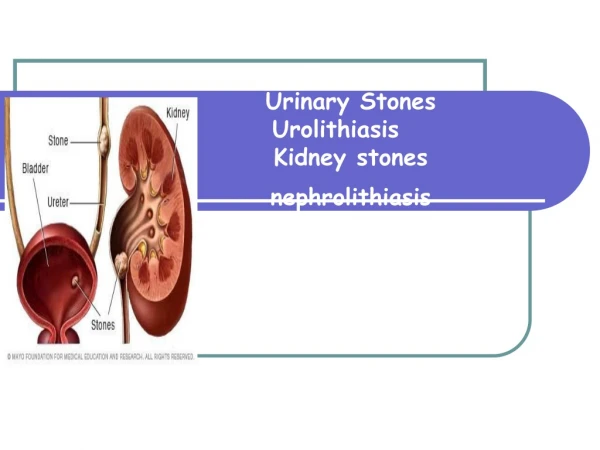
Pathophysiology • Stone formation occurred in kidney and urinary bladder. • Kidney → ureter; • bladder→ urethra
Outcomes of urinary tract calculi: • Direct injuries; • Obstruction, hydronephrosis, urinary retention; • Infection, UTI; • Malignance. squamous cell carcinoma of the renal pelvis.
Chapter 2Upper Urinary Tract Calculi Renal and ureteral calculi. • Three distinct narrowings of ureter: • 1. the ureteropelvic junction; 2. crossing of the iliac vessels; 3. the ureterovesical junction.
The ureter demonstrating sites of normal functional or anatomic narrowing at the ureteropelvic junction (UPJ), the iliac vessels, and the ureterovesical junction (UVJ).
Clinical presentation: • Hematuria, gross or microscopic hematuria, latter is more common; • dull flank pain; • renal colic, severe flank pain in the costovertebral angel area of the back and extending anteriorly and inferiorly on the abdomen, sometimes cause pain in scrotum and testes; • Symptoms of bladder irritability may be noted when ureteral stone approaches the bladder; • Nausea, vomit; • Symptoms of urinary tract infection: fever, lower urinary tract syndrome.
Physical examination: • Tenderness in the flank area on the side of involved kidney; • A palpable mass in the upper quadrant area in some patients with chronic hydronephrosis; • Nausea, vomit; • Symptoms of urinary tract infection: fever, lower urinary tract syndrome.
Diagnosis • Patient history and physical examination; • Hematuria and pain, especially renal coli after movement; • Urinalysis: hematuria, pyuria, urinary PH changes, crystals; • Differentiated diagnosis: acute appendititis, ectopic pregnancy, bile duct stone, pyelitis.
Diagnosis 1、KUB: 90% stones could be found in KUB. calcium oxalate, calcium phosphate, magnesium ammonium phosphate, cystine stones are usually radiopaque. Uric acid and xanthine stones are “negative” in KUB. 2、IVU:intravenous pyelogram may be obtained to confirm the presence of radiolucent stones and also to identify any anatomic abnormalities that may predispose the patient to stone formation. IVU is also useful in detecting the site and extent of obstruction. In patients with significant azotemia (creatinine>3mg/dl), caution should be exercised in use of IVU. 3、Sonography:non-invasive, the first-line imaging modality when renal colic is suspected in pregnancy. 4、Metabolic evaluation.
Diagnosis 4、RGP: retrograde pyelography 6、CT, to quickly image the entire collecting system in a rapid sequence, is also useful to detect non-opaque calculi as well as opaque calculi. 7、ECT 8、cystoscopy, ureteroscopy
management • Many factors should be considered in choosing the appropriate management: the size, location of the calculi, obstruction, infection, renal function.
Conservative management • Size<0.6cm,no obstruction or infection; • Pure uric acid and cystine calculi; • More than 80% of ureteral calculi with diameter less than 0.4cm will pass spontaneously; • However, small ureteral calculi remain in the same position for six weeks or longer should be considered candidates for manipulation or extraction.
Conservative management 1. Pain relief; 2. Increasing the oral intake of water: which is the primary method of reducing the saturation of urine with calculus-forming components, at least 2 to 3 liters per day; 3. Dietary Recommendations : calcium-containing food: milk, tofu, chocolate, nuts; oxalate-containing food: tea, tomato, spinage, cocoa; purine-containing food: beer, gut.
Conservative management 4. Management of infection: antiboitics; 5. Different kinds of calculi: ⑴uric acid calculus: alkalizing urine to a mean PH between 7 and 7.5, potassium citrate, sodium bicarbonate. Allopurinol is beneficial in certain cases of very active stone formation; ⑵struvite calculus: ammonium chloride, urease inhibitor acetohydroxamic acid, control of urinary tract infection; ⑶cystine calculus: increasing the urine output to 2 to 4 liters per day, raising urinary PH to 7.5, or D-penicillamine administration in severe cases.
ESWL (extracorporeal shock wave lithotripsy) • Indications: renal or ureteral calculi with diameter<2cm, no obstruction in distal urinary tract. • Contraindication:obstruction in the distal part of the urinary tract, coagulopathy, severe arrhythmia, patients with pacemaker, creatine≥265μmol/L, acute urinary tract infection, lower ureteral stones in pregnancy.
PCNL(percutaneous nephrostolithotomy) Indication: renal calculi with diameter more than 2cm or upper ureteral calculi failed to SWL.
ureteroscopic lithotomy or lithotripsy • Indication: Ureteral calculi located in middle or lower portion of ureter; failed to SWL; nonopaque ureteral calculi failed to conservative management, steinstrasse.
Laparoscopy; Open surgery: Indication: Patients failed to other mini-invasive procedure. Seldom performed recently.
Management of bilateral upper urinary tract lithiasis • 1, Bilateral ureteral calculi: Calculus accompanied with more severe obstruction should be treated first; • 2, Renal calculus with ureteral calculus on the other side, ureteral calculus should be treated first.
Prevention • General prevention: increasing oral intake of water; • Uric acid calculi; • Cystine urolithiasis; • Struvite calculi; • Correcting the existing metabolic or other causes: hyperparathroidism, urinary tract infection, urinary tract obstruction, etc.
Chapter 3Bladder Calculi • Migrant Calculi. • Primary Idiopathic (Endemic) Calculi; it is rare in developed countries, a mixed cereal diet with milk supplements reduces the incidence of endemic bladder calculi. • Secondary Bladder Calculi, These secondary bladder calculi are most often related to urinary stasis or recurrent urinary tract infection due to bladder outlet obstruction or neurogenic bladder dysfunction.
Symptoms • Some bladder calculi are asymptomatic and are found incidentally. • The typical presentation in patients with symptomatic bladder calculi includes intermittent, painful voiding and hematuria.
Diagnosis 1. Symptoms; 2. KUB; 3. Ultrasonography 4. Cystoscopy, which is the single most accurate examination to document the presence of a bladder calculus.
Radiographic appearance of uric acid bladder calculi. A, Multiple low densities are seen in the pelvis on the scout film. B, The filling defects (calculi) settle to the bladder base on the upright excretory urogram.
Radiographic (A) and gross appearance (B) of a calcium oxalate monohydrate calculus (jackstone).