Download
1 / 1
10 likes | 119 Views
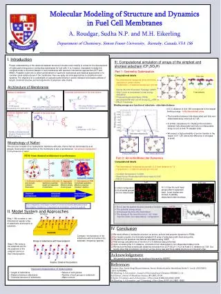
Dynamics in Modeling and Localization of ECG and EEG. Burak Erem*, Jaume Coll Font*, Ramon Martinez Orellana *, Petr Stovicek + , Dana H. Brooks* (*) CDSP Center, ECE Dept., Northeastern Univ., Boston, USA (+) Charles University Hospital, Prague, Czech Republic.

E N D
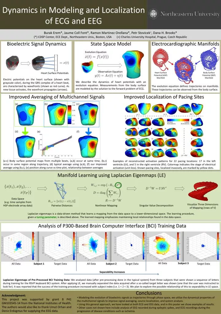
Dynamics in Modeling and Localization of ECG and EEG Burak Erem*, JaumeColl Font*, Ramon Martinez Orellana*, PetrStovicek+, Dana H. Brooks* (*) CDSP Center, ECE Dept., Northeastern Univ., Boston, USA (+) Charles University Hospital, Prague, Czech Republic Bioelectric Signal Dynamics State Space Model Electrocardiographic Manifolds Evolution Equation Observation Equation Heart Surface Potentials Heart Surface Potential (HSP) Manifold Body Surface Potential (BSP) Manifold Electric potentials on the heart surface (shown with grayscale color), during the QRS complex of a heart beat, are characterized by wavefronts (shown as red curve). As new tissue activates, the wavefront propagates (arrows). We describe the dynamics of heart potentials with an evolution equation. Measurements from the body surface are modeled by the solution to the forward problem of ECG. The evolution equation defines trajectories on manifolds. These trajectories can be observed from the body surface. Improved Localization of Pacing Sites Manifold Learning using Laplacian Eigenmaps (LE) Data Space (e.g. time samples from HSP electrode array data) Visualize Three Dimensions of Mapping (rows of V) Pairwise Distances Nonlinear Mapping Singular Value Decomposition Laplacian eigenmaps is a data-driven method that learns a mapping from the data space to a lower-dimensional space. The learning procedure, given a tuning parameter, is described above. The learned mapping emphasizes maintaining local relationships found in the data space. Improved Averaging of Multichannel Signals (a-c) Body surface potential maps from multiple beats, (a,b) occur at same time, (b,c) occur in same region along trajectory, (d) typical average using (a,b), (f) our improved average using (b,c), (e) position along curve vs time plot, relationship between averages Examples of reconstructed activation patterns for 22 pacing locations: 17 in the left ventricle (LV), and 5 in the right ventricle (RV). Colormap indicates the stage of electrical activation (unit-less). Known pacing sites, localized invasively, are marked by yellow dots. Analysis of P300-Based Brain Computer Interface (BCI) Training Data Subject 3 All Data All Data Subject 1 Target Data All Data Subject 2 Target Data Target Data SeparabilityIncreases Laplacian Eigenmaps of Pre-Processed BCI Training Data: We analyzed data (after pre-processing done in the typical system) from three subjects that were shown a sequence of letters during training for the RSVP keyboard BCI system. After applying LE, we manually separated the data acquired after a so-called target letter was shown (one that the user was instructed to look for). It was reported that the success of the training procedure increased with subject index (i.e. 1 < 2 < 3). We plan to explore the possible relationship of this to separability in LE space. Conclusions Acknowledgment: This project was supported by grant 8 P41 GM103545-14 from the National Institutes of Health. The authors would also like to thank UmutOrhan and DenizErdogmus for supplying the EEG data. • Modeling the evolution of bioelectric signals as trajectories through phase space, we utilize the dynamical properties of the multichannel signals to improve signal averaging, source localization, and system analysis. • Using this general approach, we have looked at both ECG and EEG data, and in this poster we show examples of results. • Other cases not shown here include analysis of EEG recorded during epileptic spikes, and ECG recordings during the progression of disease conditions such as ischemia.