Download
1 / 1
20 likes | 464 Views
Brain Perfusion and Blood-Brain Barrier Permeability Imaging. Max Wintermark, M.D. Director of the UCSF NeuroCardioVascular Imaging Lab. Bruno P. Soares, M.D. Senior Research Fellow

E N D
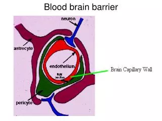
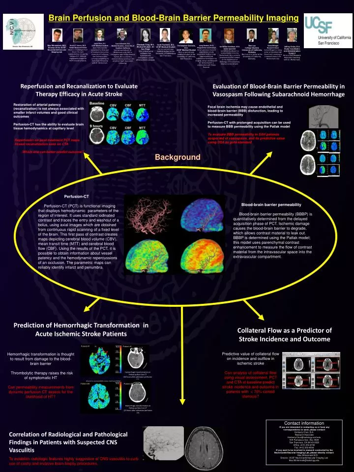
Brain Perfusion and Blood-Brain Barrier Permeability Imaging Max Wintermark, M.D. Director of the UCSF NeuroCardioVascular Imaging Lab Bruno P. Soares, M.D. Senior Research Fellow Bruno is a radiologist from Brazil. His main responsibilities include the review of CT, MRI and angiography images for NCVI research projects. Jason Hom, B.S. UCSF Medical Student Jason Hom recently completed his third year of medical school at the University of California, San Francisco. He is currently completing his Doris Duke Fellowship with Dr. Wintermark. Tom Schneider, B.S.Medical Student, University of Southern California Tom has worked with Dr. Wintermark since 2007 in conjugation with Phillips Medical System. He is currently a research scholar working in the American Heart Association Fellowship. Scott Fischette, B.S.UCSF Medical Student He is a second year medical student at the University of California at San Francisco. Sumail BhogalResearch Intern Sumail Bhogal is a rising senior at Stockdale High School and is working as a research intern in Dr. Wintermark's lab. Christopher Gouveia, B.A. UCSF Medical Student He is a second year medical student at the University of California, San Francisco. Elizabeth Tong, M.S. Medical Student, UC San Diego She is developing software that automatically characterize carotid atherosclerotic plaques. Patient #1 Patient #1 Hemorrhagic transformation on Follow-up Non-Con CT (26 hours after admission perfusion CT) Patient #2 Patient #2 Hemorrhagic transformation on Follow-up Non-Con CT (23 hours after admission perfusion CT) Joerg Bredno, Ph.D.Programmer and AnalystJoerg’s previous research experiences include automated medical image segmentation and quantitative functional x-ray imaging. Joerg is working with Dr. Wintermark in a collaboration with Philips Research. Ben Lau College Student, University of California, Berkeley Ben Lau is a Junior at the University of California, Berkeley. Jan Willem Dankbaar, M.D. Junior Specialist Jan Willem Dankbaar is a medical doctor from the Netherlands. He is currently employed as a PhD student at the Department of Radiology of the University Medical Center Utrecht. Jeffrey Chien, B.A. Study Coordinator and Administration Support He is a Staff Research Associate II working with Dr. Wintermark. Reperfusion and Recanalization to Evaluate Therapy Efficacy in Acute Stroke Evaluation of Blood-Brain Barrier Permeability in Vasospasm Following Subarachnoid Hemorrhage • Restoration of arterial patency (recanalization) is not always associated with smaller infarct volumes and good clinical outcomes • Perfusion-CT has the ability to evaluate brain tissue hemodynamics at capillary level • Reperfusion on post-treatment PCT maps • Vessel recanalization seen on CTA • Which one can better predict outcome? Focal brain ischemia may cause endothelial and blood-brain barrier (BBB) disfunction, leading to increased permeability Perfusion-CT with prolonged acquisition can be used to measure BBB permeability using the Patlak model To evaluate BBB permeability in SAH patients suspected of vasospasm, and its predictive value using DSA as gold-standard Background • Perfusion-CT • Perfusion-CT (PCT) is functional imaging that displays hemodynamic parameters of the region of interest. It uses standard iodinated contrast and traces the entry and washout of a bolus, using axial images which are obtained from continuous rapid scanning of a fixed level of the brain. This first pass of contrast creates maps depicting cerebral blood volume (CBV), mean transit time (MTT) and cerebral blood flow (CBF). Using the results of the PCT, it is possible to obtain information about vessel patency and the hemodynamic repercussions of an occlusion. The parametric maps can reliably identify infarct and penumbra. • Blood-brain barrier permeability • Blood-brain barrier permeability (BBBP) is quantitatively determined from the delayed acquisition phase of PCT. Ischemic damage causes the blood-brain barrier to degrade, which allows contrast material to leak out. BBBP is determined using the Patlak model; this model uses parenchymal contrast enhancement to measure the flow of contrast material from the intravascular space into the extravascular compartment. Prediction of Hemorrhagic Transformation in Acute Ischemic Stroke Patients Collateral Flow as a Predictor of Stroke Incidence and Outcome Hemorrhagic transformation is thought to result from damage to the blood-brain barrier Thrombolytic therapy raises the risk of symptomatic HT Can permeability measurements from dynamic perfusion-CT assess for the likelihood of HT? Predictive value of collateral flow on incidence and outflow in ischemic stroke Can analysis of collateral flow using visual assessment, PCT and CTA at baseline predict stroke incidence and outcome in patients with < 70% carotid stenosis? Absolute permeability maps (ml/min/100g) from admission perfusion-CT scans • Contact information • If you are interested in contacting us or have any • correspondence to send, please contact:Kimberly Chun, B.A.Research AssociateKimberly.Chun@radiology.ucsf.edu505 Parnassus Ave., Box 0628San Francisco, CA 94143-0628 Office: (415) 353-8749Fax: (415) 353-8593 • If you want to be involved in research conducted by the NeuroCardioVascular Imaging Lab, please directly contact: Max Wintermark M.D.Director, UCSF NeuroCardioVascular Imaging Lab Max.Wintermark@radiology.edu Correlation of Radiological and Pathological Findings in Patients with Suspected CNS Vasculitis To establish radiologic features highly suggestive of CNS vasculitis to curb use of costly and invasive brain biopsy procedures.