Download
1 / 23
240 likes | 335 Views
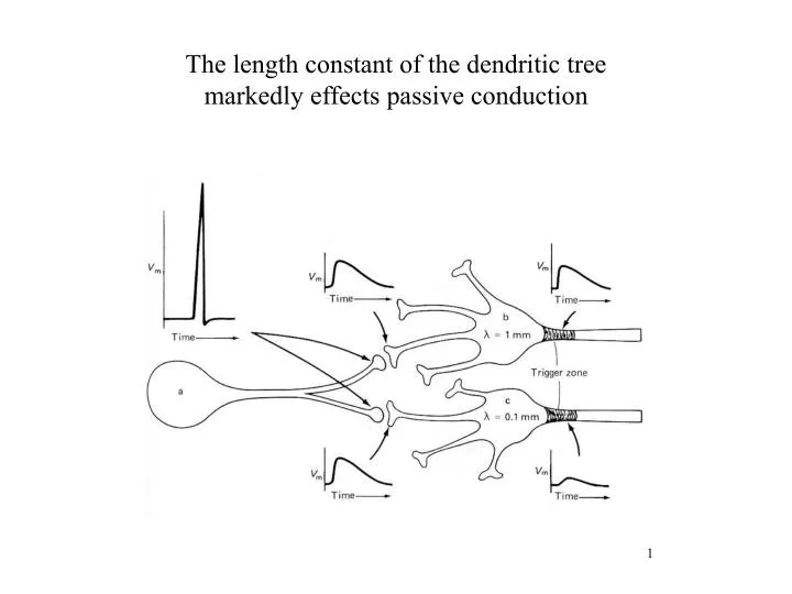
The length constant of the dendritic tree markedly effects passive conduction.

E N D
The length constant of the dendritic tree markedly effects passive conduction
Diversity in dendritic excitability. All recordings are from different neocortical layer 5 pyramidal neurons, with the recording locations indicated schematically on a typical morphology. (Left) Action potential evoked by distal synaptic stimulation recorded simultaneously in the axon (green trace), soma (blue trace), and apical dendrite 300 µm from the soma (orange trace). The action potential is initiated in the axon and backpropagates into the soma and dendritic tree [modified from (63)]. (Right) Initiation of dendritic Na+ and Ca2+ spikes. In each case, recordings were made simultaneously from the soma and distal dendrites (400 µm from the soma for the Na+ spike, orange trace; 920 µm from the soma for the Ca2+ spike, red trace), and both a subthreshold and a suprathreshold response to distal synaptic activation are shown
Diversity of compartmentalization in dendrites caused by cell-specific differences in dendritic morphology. Action potential backpropagation and forward propagation of a dendritic spike was simulated in a neocortical layer 5 neocortical pyramidal neuron (A and C) and a cerebellar Purkinje cell (B and D). Both morphologies were provided with the same uniform dendritic density of voltage-gated Na+ and K+ channels [35 and 30 pS/µm2, respectively to isolate the effect of dendritic morphology. The local amplitude of the backpropagating action potential (AP) or dendritic spike is coded by color in each cell type.
Modulation of dendritic excitability and interactions between electrical compartments. (A) Dynamic compartmentalization of dendrites by inhibition in an olfactory bulb mitral cell. Activation of an inhibitory postsynaptic potential (IPSP) prevents forward propagation of a dendritic spike to the soma. (B) Activity-dependent backpropagation of action potentials in a hippocampal CA1 pyramidal neuron. Action potentials evoked by a somatic current pulse decline progressively in the distal dendrites. (C) Modulation of action potential backpropagation by subthreshold oscillations in dendritic membrane potential in a neocortical layer 5 pyramidal neuron. (D) Pairing a backpropagating action potential (AP) with distal EPSPs (simulated by dendritic current injection) in a neocortical layer 5 pyramidal neuron leads to a distal Ca2+ spike and a somatic burst.
Chemical computation and compartmentalization in dendrites. (A) Chemical compartmentalization in cerebellar Purkinje cell dendrites. Local dendritic Ca2+ signals (F/F) in response to synaptic stimulation of parallel fibers (PF, left) or uncaging of IP3. (B) Coincidence detection in dendritic spines mediated by supralinear Ca2+ signals. Line scan (left) and Ca2+ signals (F/F, right) in spines of a hippocampal CA1 pyramidal neuron in response to conjunction of synaptic input (SY) and a train of backpropagating action potentials (AP). The synaptic current in the whole-cell recording (Iwc) is shown below. (C) In vivo measurements of dendritic Ca2+ dynamics in neocortical layer 5 neocortical pyramidal neurons. Top: Schematic illustration of the recording arrangement, which also allows stimulation of synaptic input (bipolar) and recording of the electrocorticogram (EcoG). Bottom: Fluorescence changes in the apical tuft in response to backpropagating action potentials and Ca2+ spikes associated with a somatic action potential burst.
Some receptors - e.g. the NMDA receptors are good examples of further complexity in ligand-gated channel function.
Oscillations can be activated in spinal cord neurons which require NMDA receptor activation
The NMDA receptor is Ca permeant, and Ca oscillates during these TTX resistant oscillations in membrane potential.