Download
1 / 35
590 likes | 1.92k Views
Tear Film, Lacrimal and Meibomian Glands. Ladan Espandar, MD, MS Assistant Professor of Ophthalmology Department of Ophthalmology University of Pittsburgh School of Medicine. DIAGRAM OF A HUMAN EYE. Sagittal section through the ocular surface. Anterior Chamber.

E N D
Tear Film, Lacrimal and Meibomian Glands Ladan Espandar, MD, MS Assistant Professor of Ophthalmology Department of Ophthalmology University of Pittsburgh School of Medicine
Sagittal section through the ocular surface Anterior Chamber The ocular surface epithelium is continuous (pink) with regional specializations in the cornea, conjunctiva, lacrimal, and accessory lacrimal glands, and meibomian gland. Each specialized region of this ocular surface epithelium contributes components of the tear film (blue).
Frontal view of the Ocular Surface System The ocular surface includes the surface and glandular epithelia of the cornea, conjunctiva, lacrimal gland, accessory lacrimal glands, and meibomian gland. The functions of the system’s components are integrated or linked by innervation, and the endocrine, vascular, and immune systems
Tear Film • The tear film is a complex composite with multiple sources, which include the lacrimal gland, meibomian glands, goblet cells, and accessory lacrimal glands (Krause, and Wolfring). • The function of the tear film includes lubrication, protection, nutrition of the cornea, and a critical role in the optical properties of the eye • Normal tear volume:6 μL • Production rate: 1.2 μL/minute • turnover rate: 16% per minute
Lipid Layer of Tear Film • The meibomian glands are the main source of lipids for the human TF. • The meibomian gland secretions: extremely complex mixture of various polar and nonpolar lipids containing cholesteryl esters (CEs), triacylglycerol, free cholesterol, free fatty acids (FFAs), phospholipids, wax esters (WEs), and diesters.
Lipid Synthesis in Meibomian Glands • Oleic acid is the most abundant fatty acid, 18C, monounsaturated (18:1(cis#9) • Key enzymes: Fatty acid and cholesterol synthesis enzymes. • Regulatory mechanism: sex steroids, corticosteroids, hypothalamic and pituitary hormones, insulin, retinoids, thyroxine, melanocortins, neurotransmitters, growth factors, and peroxisome proliferator activated receptor ligands. Wax ester
Embryologic Development of theMeibomian Gland • Third to the seventh month of gestation, during the sealed lid phase of eyelid development. • Loose connective tissue of the mesoderm in the lid folds differentiates into the tarsal plate and muscles (orbicularis and Riolan’s muscle), the blood vessels, and the loose connective tissue underlying the outer lid skin and the conjunctiva. • Meibomian gland anlage grow from the ectodermal sheet
Histologic Appearance of the Meibomian Gland (A) The holocrine acini filled with the secretory cells (meibocytes) and surrounded by a basement membrane (bm). (B) In the area of the disintegration zone, located at the transition of the acinus to the ductule (C) Numerous acini of spherical to elongated shape are radially arranged around the central duct (cd) of a gland, seen here in a longitudinal section
Physiology of the Meibomian Glands The driving forces (1) the continuous secretion of meibum by the secretory acini (2) the mechanical muscular action by muscle fibers of the pretarsal orbicularis muscle (M. orbicularis), and of the marginal muscle of Riolan (M. Riolan), which encircles the terminal part of the meibomian gland. During a blink, these muscles may exertca compression of the meibomian gland that drives the oil out of the orifice into the marginal lipid reservoir, where it eventually constitutes the tear film lipid layer (TFLL), as observed clinically
MEIBOMIAN GLAND STEM CELLS ANDCELL DYNAMICS • After a process of maturation including lipid synthesis and accumulation, centripetal cell movement, and eventual cell degeneration and membrane disintegration, the lipids and other cell components are shed into the lumen of the ductal system. • This holocrine secretion process hence results in the dynamic consequence that secretory cells are continuously lost and replaced. • The continuous loss of acinar cells requires a consequent continuous production of new cells and therefore a continuous cell turnover and differentiation within the acinus. • Meibomian Gland Stem Cells concluded from their observations that the stem cells of the meibomian glands lie at the circumference of each acinus
Innervation • The meibomian glands of the human have a dense meshwork of unmyelinated nerve fibers (nerve plexus) around the acini outside the basement • Mainly represent cholinergic parasympathetic nervous system, from the pterygopalatine ganglion, sympathetic nerves from the superior cervical ganglion and sensory fibers from the trigeminal ganglion. • Other neuropeptides: calcitonin gene-related peptide (CGRP) and substance P, vasoactive intestinal polypeptide.
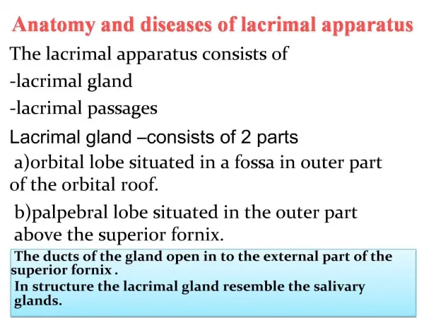
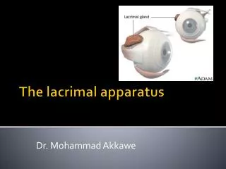
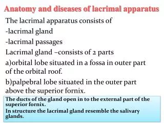
ANATOMY OF THE HUMAN LACRIMAL GLAND Main lacrimal gland, accessory lacrimal gland. The main lacrimal gland: palpebral and orbital lobes, separated by aponeurosis of levator palpebrae superioris Excretory ducts coming from the palpebral and orbital lobes open into the superior conjunctival fornix. The accessory lacrimal gland is divided into 2 anatomic groups: the glands of Krause and glands of Wolfring. The glands of Krause are located in the lamina propria of fornix and the glands of Wolfring are in the edge of the tarsus. The ducts of both glands open on the conjunctival surface.
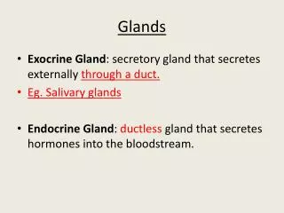
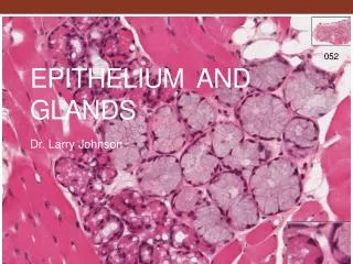
HISTOLOGY OF THE HUMAN MAIN LACRIMAL GLAND The lacrimal gland is an exocrine gland. The main lacrimal gland comprises many lobules separated from one another by loose connective tissue. Each lobule has many acini and intralobular ducts. The connective tissue contains interlobular ducts, vessels, nerve fibers, fibroblasts, many plasma cells, and a few lymphocytes. Interlobular ducts finally become approximately 12 excretory ducts, which open into the fornix of conjunctiva.
Histology The acinus comprises pyramid-shaped acinar cells with central lumen. Acinar cells have many periodic acid-Schiff (PAS)-positive secretory granules and basally located nuclei. Myoepithelial cells are distributed surrounding the acini and intercalated ducts. Myoepithelial cells have stellate, multiprocessed morphology, and their contraction may play a role in expelling secretory products from glandular lumina into ducts. Plasma cells in connective tissue produce IgA and are involved in the formation of secretory IgA in tear fluid. Secretory IgA is the best-defined effector component of the mucosal immune system.
HISTOPATHOLOGY OF THE HUMAN MAIN LACRIMAL GLAND Acinar atrophy; periacinar fibrosis; periductal fibrosis; interlobular ductal dilatation; interlobular ductal proliferation; lymphocytic infiltration; and fatty infiltration. Chronic graft-versus-host disease (GVHD), stromal fibroblasts are actively involved in the pathogenic process of the lacrimal gland. In Sjögren syndrome, the earliest histologic finding in salivary glands has been described as periductal lymphocytic infiltration.
Aqueous Tear Components • Contains proteins, and electrolytes • 6–10 mg/mL total proteins and almost 500 proteins • Major tear proteins include lysozyme, lactoferrin, secretory immunoglobulin A (sIgA), serum albumin, lipocalin (previously called tear-specific prealbumin), and lipophilin. • Chloride and potassium are higher in the tears (tears, 120 mEq/L and 20 mEq/L; serum, 102 mEq/L and 5 mEq/L, respectively) • Glucose concentration is lower in the tears (about 2.5 mg/100 mL) compared to plasma (85 mg/L). • The osmotic pressure of the tears ranges between 280 and 305 mOsm/L. • Tear proteins vary with the state of health of the ocular surface.
Comparison of Tear Components with that of Serum
Conjunctiva • The conjunctiva is a mucous membrane that protects the soft tissues of the eyelid and orbit, allows extensive movement of the eye and is the main site for the production of the aqueous and mucous components of tears. Embryology • The conjunctiva develop within the lid folds from surface ectodermal and neural crest tissue along the posterior surface of the lids and from similar tissues around the developing cornea.
Histology The conjunctival surface is composed of stratified nonkeratinizing squamous epithelium The epithelium contains goblet cells, Langerhans cells, and dendritic melanocytes. The substantiapropria, or conjunctival stroma, is highly vascularized and may contain nonstriated muscle, sympathetic nerves, and fatty tissue. Apically, a glycocalyx is secreted from mucin-containing intraepithelial vesicles, consisting of transmembranemucins (MUC1, MUC4, MUC16). The long-chain glycoprotein molecules maintain tear film stability by anchoring the mucin produced by the goblet cells (MUC5AC) to the conjunctival surface and also bind to immunoglobulins. Lymphocytes, dendritic melanocytes, and Langerhans cells may be seen in the suprabasal region of the epithelium.
Glycocalyx Muc 16 (TEM) The cytoplasmic tail of MUC16 is tethered to the actin cytoskeleton through actin linking ERM family of proteins (ezrin, rodixin moesin, and merlin). • Mat of long chain glycoproteins integral to the superficial corneal epithelial cell membranes. • Helps create a hydrophilic surface on the hydrophobic cell membranes and protects against bacterial pathogens • Insufficient mucins can damage glycocalyx, causing the tear film to destabilize and break up before a blink can occur, exposing the cornea to air and pathogens H185 carbohydrate epitope of Muc16 (TEM) Muc 16 (SEM) SEM showing corneal surface microplicae
MUC16 Facilitates the Ocular Surface Barrier Function • siRNA knockdown of mucin-16 in a human corneal limbal epithelial cell line. HCLE cells cultured to subconfluent or confluent stages do not express MUC16, and rose bengal dye penetrates all cells. After the cells are cultured seven additional days, cells stratify and express MUC16. Islands of differentiated cells exclude the Rose bengal dye. When MUC16 was knocked down by 80% to 90%, the islands that exclude the dye were diminished. • (B) Rose bengal stains areas of damaged ocular surface epithelium in dry eye
Conjunctival goblet cells Goblet cells are unicellular, mucin-secreting glands that account for approximately 5% to 10% of basal cells. They are likely apocrine in nature, with all the secretory granules secreted once the cell has been activated. They are the primary source of the large soluble mucins in the tear film. Goblet cells release their secretory granules in response to activation of the parasympathetic nerves that surround them. Sympathetic nerves also surround the goblet cells, not sensory. The mucin synthesized by goblet cells of normal human conjunctiva is identified as MUC5AC Goblet cells may also secrete MUC1, MUC4, and MUC16. These mucins function to protect, hydrate, and lubricate the ocular surface.
Maintenance of tear film stability Production and secretion of tear components, redistribution and drainage of the tears are precisely regulated
Control of Tear Secretion The small sensory nerve endings located just below the epithelial surface of the cornea, lid margin, and conjunctiva constantly respond to drying and temperature change as well as contact and chemical changes, by sending intensity-coded neural signals to the spinal trigeminal nucleus located in the brain stem. A multisynaptic pathway to the preganglionic parasympathetic nuclei in the superior salivatory nucleus forms the output to the secretory tissue.
Dry Eye 2007 International Dry Eye Workshop Dry eye is a multifactorial disease of the tears and ocular surface that results in symptoms of discomfort, visual disturbance, and tear film instability with potential damage to the ocular surface. Dry eye is accompanied by increased osmolarity of the tear film and inflammation of the ocular surface. • Tear osmolarity increases by an average of ~13% • HLA ClassII expression by conjunctival epithelial cells increases • CD4+ T-Cells infiltrate conjunctiva • Tear MMP-9 activity and expression of cytokines increases • Lacrimal proteins and conjunctival goblet cell density decreases
Major etiological causes of dry eye Report of the International Dry Eye Workshop. The Ocular Surface. 2007. 5(2) 69-201
Mechanisms of dry eye Report of the International Dry Eye Workshop. The Ocular Surface. 2007. 5(2) 69-201
SUMMARY • Ocular surface consists of the cornea, bulbar conjunctiva and the palpebral conjunctiva, bathed in the tear film. • Tear film consists of the anterior lipid layer (secreted by the meibomian glands), central aqueous layer (secreted by the lacrimal glands) containing mucins (secreted by conjunctival goblet cells), anti-bacterial peptides, growth factors, antibodies, solutes, and the posterior glycocalyx (superficial epithelial cell membrane bound mucins). • Dry eye is a widely prevalent multifactorial disorder of the break up of the tear film.