Download
1 / 39
410 likes | 431 Views
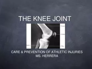
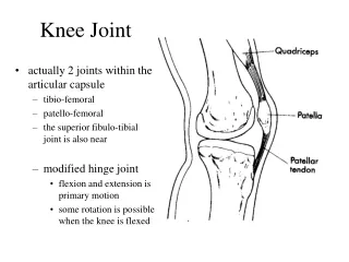
knee joint, muscles and actions of the knee. Semmelweis Egyetem ÁOK Anatómiai Intézet Dr. Csáki Ágnes 2017.10.25. The knee joint is the largest synovial joint in the body. It consists of: two articulation between the femur and tibia (condylar joint) , which is weight bearing;

E N D
knee joint, muscles and actions of the knee Semmelweis Egyetem ÁOK Anatómiai Intézet Dr. Csáki Ágnes 2017.10.25.
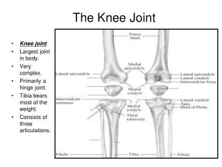
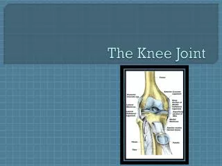
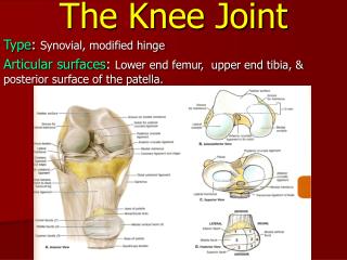
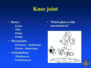
The knee joint is the largest synovial joint in the body. It consists of: two articulation between the femur and tibia (condylar joint), which is weight bearing; the articulation between the patella and the femur. Fibula is not directly involved in the joint.
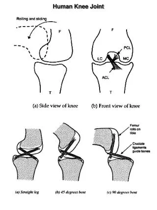
Tibiofemoral and patellofemoral joints Mechanism: trochoginglymus !!! Originally two joints: 4 collateral ligaments! (lateral medial collaterals and anterior and posterior crutiate) Incongruentsurfaces: equalized by menisci Great workload, frequently damaged, especially the ligaments and the menisci. Movements: flexion:130° ; extension:0-5° ; rotation:50° only at flexed knee!!! osseous structures, extraarticular structures, and intraarticular structures
FEMUR the distal end of femur is characterized by two large condyles, which articulate with the proximal end of the tibia. The condyles are separated posteriorly by an intercondylar fossa and are joined anteriorly with patellar surface. There are bony elevations on the nonarticular outer surfaces of the condyles - lateral and medial epicondyles, which serve for attachment of collateral ligaments of the knee joint. The articular surface of the medial condyle is longer than that of the lateral condyle, but the lateral condyle is wider. The long axis of the lateral condyle is oriented essentially along the sagittal plane, whereas the medial condyle turns medially
TIBIA The expanded proximal end of the tibia forms two rather flat surfaces, condyles. They are separated in the midline by the intercondylar eminence with its medial and lateral intercondylar tubercles. Anterior and posterior to the intercondylar eminence are the areas that serve as attachment sites for the cruciate ligaments and menisci. Intercondylar area
PATELLA (knee cap) is the largest sesamoid bone in the body triangular: its apex is pointed inferiorly for attachment to the patellar ligament, its base is broad and thick for the attachment of the quadriceps femoris muscle from above; its posterior surface articulates with the femur andis divided by a vertical ridge, resulting in a smaller medial and a larger lateral articular facet. The patella is anchored and stabilized to the knee by four structures : the patellar tendon inferiorly, the quadriceps tendon superiorly , and the patellar retinaculum medially and laterally. (attachement of quadriceps straight to the tibia)
Functionsprimarilyas an anatomicpulleyforthequadricepsmuscle. Itincreasesthe lever arm of theextensormechanismallowingfor more effectivekneeextension and thusincreasequadricepsstrengthby 33–50%. itactsas a “spacer” and protectsthetendonbyreducingthefrictionandcompressivestressandminimizestheconcentration of stressbytransmittingforcesevenlytotheunderlyingbone Patellar dislocation
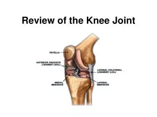
Ligaments Extracapsular Patellar ligament Medial collateral lig. Lateral collateral lig. Oblique popliteal lig. Intracapsular Medial and lateral cruciate lig. Meniscofemoral lig. Transverse lig. of knee
Tibial (medial) collateral ligament – A wide and flat ligament, found on the medial side of the joint. Proximally, it attaches to the medial epicondyle of the femur, distally it attaches to the medial surface of the tibia. Reinforces the capsule, attaches to the meniscus Fibular (lateral) collateral ligament – Thinner and rounder than the tibial collateral, this attaches proximally to the lateral epicondyle of the femur, distally it attaches to the lateral surface of the fibular head.
under the fibular (lateral) collateral ligament – runs the tendon of popliteal muscle so the ligament does not attach to the capsule
Collateral ligaments Streched and therefore work only at extended knee. If the joint is flexed to at least 30 degrees, they do not limit any movements (rotation is possible). Test at extended knee (try to open the joint medially or laterally). (At a flexion more than 30 degrees the cruciate ligaments take over the same functions.) Sobota - Atlas of Human Anatomy
Oblique popliteal ligament reinforces the posterior capsule, attachment of the semimembranous muscle Arcuate popliteal ligament reinforces of the fibrous capsule above the popliteal muscle
Cruciate ligaments Anterior and posterior crutiate ligaments are streched between the internal surface of the condyles of the femur and intercondylar areas of the tibia The cruciate ligaments function as stabilizers of the joint and axes around which rotary motion, both normal and abnormal, occurs. They restrict the backward and forward motion of the tibia on the femur and assist in the control of both medial and lateral rotation. External rotation of the tibia produces an unwinding of the ligaments, and internal rotation produces a winding up of the cruciate ligaments
Cruciate ligaments Some parts are streched at every position, with greater workload at flexed knee – higher chance of damage (skiing). Limit medial and lateral opening, rotation, antero-posterior movements. Test: drawer sign. Anterior drawer sign: ant. cruciate lig. injury. Posterior drawer sign: post. cruciate lig. injury Sobota - Atlas of Human Anatomy
Intracapsular but extrasynovial position of the cruciate ligaments Stratum synoviale Stratumfibrosum Superior view of the tibia
Menisci The meniscus serves as a shock-absorption system, assists in lubricating the knee joint, and limits the ability to flex and extend the joint. Incongruent surfaces. Compensates the differences in surfaces Fixed but also mobile structures, obtain different positions at different stages of movements. Medial meniscus (C-shaped) is more fixed by the capsule – damaged more often. Lateral meniscus- nearly round, not fixed by the capsule –popliteal tendon! Symptoms: pain, „stop of movement” Sobota - Atlas of Human Anatomy M L Passive movements of menisci
Intraarticular structures Attachement of menisci to the intercondylar areas and eminences Transverse genicular ligament Post and ant meniscofemoral ligaments
Meniscal tear Is most commonly caused by twisting or over-flexing the knee joint. http://www.nlm.nih.gov/medlineplus/ency/article/001071.htm Arthroscopic removal of damaged part: in case of central injury
Arthroscopy Removal does not preclude normal motion, but -increase wear on articulating surfaces -increase chance of developing degenerative joint disease
The synovial membrane and bursae Atteches to the margins of the articular surfaces and to to the menisci Does not contain the crutiate ligaments Suprapatellar bursa opens to the cavity Prepatellar bursa Subcutan and deep infrapatellar bursa Infrapatellar fat pad
A Baker's cyst (popliteal cyst) is located behind the knee and is a swelling of the popliteal bursa. A Baker's cyst can form when joint-lubricating fluid fills a cushioning pouch at the back of your knee.
Genicular arteries A. descendens genus Superior lateral and medial genicular a. Inferior lateral and medial genicular a. Middle genicular a. from the popliteal a. and descending genicular a. from the femoral a.
Angle of inclination acts on the weight-bearing line of the leg Normal Coxa valga Coxa vara Genu varum Genu valgum
A child’s knee Growth cartilage or epiphyseal plate
Prosthesis of cartilaginous surface Cartilage (in joint) does not regenerate!
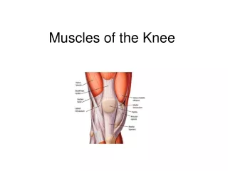
Muscles around the knee The important extraarticular structures supporting and influencing the function of this joint are the synovium, capsule, collateral ligaments, and musculotendinous units that span the joint. The musculotendinous units are principally -the quadriceps m. and sartorius m. -the hamstring muscles, -the gastrocnemius, -the popliteus, -and the iliotibial band.
quadriceps m. and sartorius m. Sartorius m. Vastus intermedius m.. Rectus femoris Vastus lateralis Vastus medialis
hamstring muscles Tensor fasciae latae m. Iliotibial tract! (Fasca lata) The four muscles of the harmstring insert below the knee They start at the ischial tuberosity, but short head of biceps from the femur. They cross the knee joint and end at the lower leg. The hamstring muscle group extend the hip and bend the knee. The harmstring muscles are the antagonists of the quadriceps m.
lateral side: insertion of the biceps femoris on the head of the fibula Biceps femoris medial side: harmstring muscles and pes anserinus
Insertion of the semitendinous m. to the pes anserinus on the medial side of he tibia together the gracilis (adductors of the tigh) and sartorius m. (extensors of tigh) „goose leg”
Three insertion of the semimembranous m.: on the medial condyle of the tibia on the fascia of the popliteus m. oblique popliteal ligament (deep pes anserinus) Origin of the gastrocnemius muscles: (the calf muscle) from the epicondyles of the femur: bending of the knee Bursae!! Origin of popliteus m.: above the soleal line from the tibia
Knee movements flexion: biceps femoris m., semitendinous m., semimembranous m., popliteus m., gastrocnemius m. extension: quadriceps femoris m. (rectus femoris m. flexes the hip joint!) medial rotation: semimembranous and semitendinous m. lateral rotation: biceps femoris m. sartorius m.: flexion of the hip, medial rotation and flexion of the knee, but near the full extension can help the extension
Literature Szentagothai J, Réthelyi M: Funkcionális anatómia, Medicina, 1989 Sobota - Atlas of Human Anatomy, 20th edition, Urban and Schwarzenberger, 1993 Renner Antal: Traumatológia, 2nd edition, Medicina, Budapest, 2003 Vízkelety Tibor: Az ortopédia tankönyve, 2nd edition, Semmelweis Kiadó, 1999 Radiologic images: http://rad.usuhs.mil/medpix/index.html