Download
1 / 33
330 likes | 345 Views
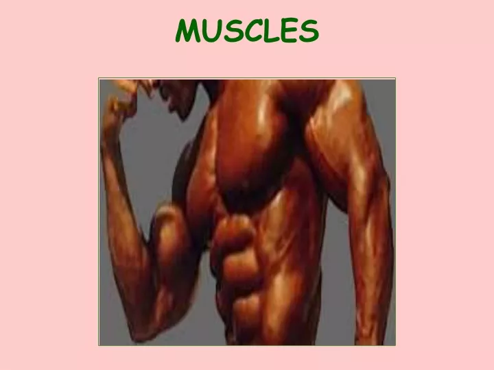
MUSCLES. I. GENERAL INFORMATION. HOW MUSCLES ARE NAMED LOCATION Ex: TEMPORALIS NUMBER OF ORIGINS Ex: BICEPS BRACHII & TRICEPS BRACHII SIZE Ex: GLUTEUS MAXIMUS & ADDUCTOR MAGNUS. LOCATION OF ORIGIN & INSERTION Ex: STERNOCLEIDOMASTOID DIRECTION OF FIBERS

E N D
I. GENERAL INFORMATION HOW MUSCLES ARE NAMED LOCATION Ex: TEMPORALIS NUMBER OF ORIGINS Ex: BICEPS BRACHII & TRICEPS BRACHII SIZE Ex: GLUTEUS MAXIMUS & ADDUCTOR MAGNUS
LOCATION OF ORIGIN & INSERTION Ex: STERNOCLEIDOMASTOID DIRECTION OF FIBERS Ex: EXTERNAL & INTERNAL OBLIQUE SHAPE OF MUSCLE Ex: DELTOID & TRAPEZIUS ACTION OF THE MUSCLE Ex: LEVATOR SCAPULAE
TERMS ACTION: WHAT A MUSCLE DOES ORIGIN: THE END OF THE MUSCLE ATTACHED TO THE BONE THAT DOES NOT MOVE INSERTION: THE END OF THE MUSCLE ATTACHED TO THE BONE THAT DOES MOVE ANTAGONISTS: MUSCLES WITH OPPOSING EFFECTS E.g.. BICEPS & TRICEPS SYNERGISTS: MUSCLES WITH THE SAME ACTION
TYPES OF MUSCLE TISSUE SKELETAL CARDIAC SMOOTH
SKELETAL MUSCLE ATTACHED TO BONES HAS STRIATIONS (stripes) VOLUNTARY CONTRACTS RAPIDLY
WALLS OF HOLLOW ORGANS • NO STRIATIONS • INVOLUNTARY
II. Muscle Tissue In Detail • OVERVIEW: • (you need about nine lines…..)
Actin Troponin Tropomyosin Myosin
II. Muscle Tissue In Detail • Skeletal Muscle Outside covering = Fascia (at muscle end may form into tendon to attach to the bone or it may form into flat sheets to connect to other muscles. These sheets are called aponeuroses.)
Next layer = epimysium (CT – connective tissue) • Wrapping fasicle (fascicle is a bundle of cells) = perimysium (CT) • Wrapping each muscle fiber (cell) in the fasicle = endomysium (CT) (muscle fiber = muscle cell)
III. MUSCLE TISSUE • Contributes to homeostasis by producing heat to maintain normal body temperature, storing and moving substances throughout the body and producing movement.
Functions • 1. Body movement • 2. Stabilizing body position • 3. Store substances like Ca2+, ATP, • 4. Move substances (ex. Food) • 5. Heat production – by muscle contractions called thermogenesis • (skip to contraction)
Properties • 1. Electric Excitability - muscle cells (and nerve cells) have the ability to respond to certain stimuli by producing electrical signals. Signals are called action potential. • 2. Contractility - the ability of muscle to contract • 3. Extensibility - the ability to stretch 4. Elasticity - ability to return to the original shape
Anatomy of a Muscle Fiber (cell) • 10 cm to 30 cm (4 in to 12 in) • 1. Has 100 + nuclei • 2. Has numerous mitochondria • 3. The number of skeletal muscle fibers is set at birth (they enlarge) • 4. Sarcolemma - (flesh sheath) plasma membrane • 5. Sarcoplasm - cytoplasm • 6. Sarcoplasmic reticulum - encircles myofibrils it stores Ca2+
Anatomy of Muscle Con’t • 7. Myoglobin - red protein only in muscles - binds O2 and releases it when the mitochondria needs it for ATP production • 8. Myofibrils- are within the fibers (cells) in the sarcoplasm. They are protein filaments that are the contractile elements/ parts of the muscle. They are striated and run the length of the muscle fiber.
Anatomy of Muscle Con’t • 9. Myofilaments - bundles in myofibrils - protein filaments made up of primarily actin (protein) and myosin (protein) • a. Thick Filaments - made up of myosin (motor protein for pushing and pulling) • b. Thin Filaments - made up of actin primarily. Also, troponin and tropormysin, which are proteins. • c. These filaments are arranged in compartments called sarcomeres (mere = parts)
Anatomy of Muscle Con’t 10. sarcomere - a contractile unit composed of thick and thin filaments
Anatomy of a Sarcomere • 1.Zone of Overlap = Thick and Thin • 2.A Band = mostly thick filaments • 3.I Band = mostly thin filaments • 4.M Line= Thickened because more myosin 5. Z Disk = Anchoring site for thin filaments 6. H Zone – only Thick filaments (myosin) P. 288
Muscle Contraction (Sliding filament mechanism) Page 295 Table 9.1 Quiz tomorrow!!!!! Contraction • A nerve impulse travels down a motor neuron axon • The motor neuron terminal releases the neurotransmitter acetylcholine (Ach) 3. AChbinds to Ach receptors 4. The carcolemma is stimulated, and a muscle impulse travels over the surface of the muscle fiber and deep into the fiber through the transverse tubules. • The muscle impulse reaches the sarcoplasmic reticulum, and calcium channels open. • Calcium ions diffuse from the sacroplasmic reticulum into the sarcoplasm and bind to troponin molecules • Tropomyosinmolecules move and expose specific sites on actin • Actin and myosin form linkages • Thin (actin) filaments are pulled toward the center of the sarcomere by myosin cross-bridges. • The muscle fiber shortens and contracts.
Muscle fiber Relaxation • Acetylcholinesterasedecomposes acetylcholine, and the muscle fiber membrane is no longer stimulated. • Calcium ions are actively transported into the sarcoplasmic reticulum. • ATP breaks linkages between actin and myosin filaments without breakdown of the ATP itself. • Breakdown of ATP “cocks” the cross-bridges. • Troponin and tropomyosin molecules inhibit the interaction between myosin and actin filaments. • Muscle fiber remains relaxed, yet ready until stimulated again.
So What is really happening? This is called the sliding filament mechanism (Theory): • Remember the sarcomere is the “functional” unit of muscle contractions • (draw sarcomere)
Review: The sarcomere shortens not the filaments! Two additional items: 1. ATPase – catalyzes the breakdown of ATP to ADP and P 2. Acetylcholinesterase – catalyzes the breakdown of ACh in the synapsis so no more Ca2+ is released from the SR.
http://www.getbodysmart.com/ap/muscletissue/contraction/contractioncycle/tutorial.htmlhttp://www.getbodysmart.com/ap/muscletissue/contraction/contractioncycle/tutorial.html • Shows contraction