Download
1 / 1
10 likes | 209 Views

E N D
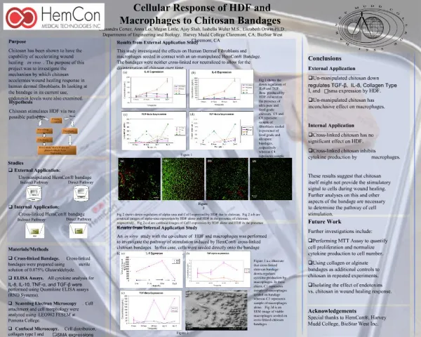
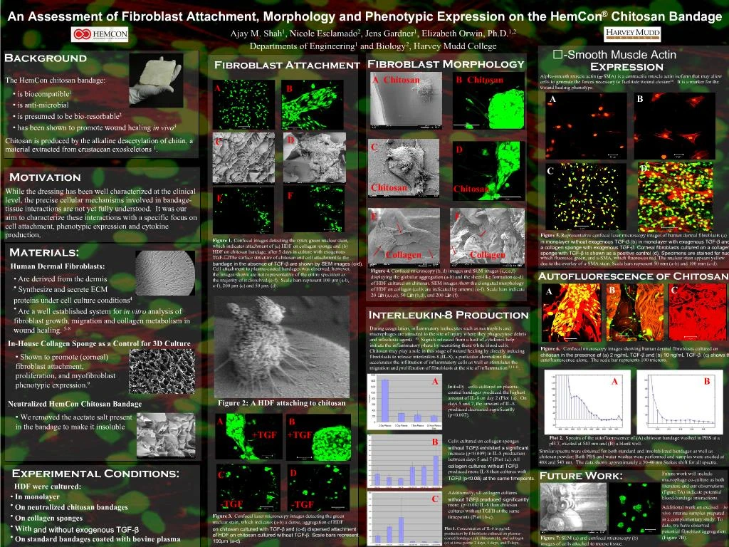
1. An Assessment of Fibroblast Attachment, Morphology and Phenotypic Expression on the HemCon� Chitosan Bandage Referencing is inconsistent
This is more readable overall than the first one (I lookeda t it too) I would fade out the background pic a little more
In the figure legends you refer to fig 1a, b etc � you need to label the pics accordingly.
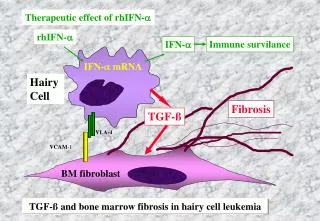
Figure 2 +/- TGF beta stuff is confusing � the point of this is that we see a confirmation in structure in SEM and confocal?
In figure 3 you start talking about stuff � tgf beta, plasma coated bandages which have not been discussed. Maybe you you lay out the experimental design somewhere � instead of methods section (which isn�t very helpful??)
Two fig on bottom of middle column are not explained � just hanging out!
Grapsh on IL8 stuff are not big enough to be readable at 100%
Table on autofluorescence stuff is not readable � take it out? Just report what we saw?
You need a box around the mouse samples too
You need a place for conclusions
You have refs which are not on there � for space considerations, make a separate sheet with refs and pin it up next to poster.
In general, this should focus on figs and not text. Figs should be bigger. Maybe get rid of legends � just put main points beneath each picReferencing is inconsistent
This is more readable overall than the first one (I lookeda t it too) I would fade out the background pic a little more
In the figure legends you refer to fig 1a, b etc � you need to label the pics accordingly.
Figure 2 +/- TGF beta stuff is confusing � the point of this is that we see a confirmation in structure in SEM and confocal?
In figure 3 you start talking about stuff � tgf beta, plasma coated bandages which have not been discussed. Maybe you you lay out the experimental design somewhere � instead of methods section (which isn�t very helpful??)
Two fig on bottom of middle column are not explained � just hanging out!
Grapsh on IL8 stuff are not big enough to be readable at 100%
Table on autofluorescence stuff is not readable � take it out? Just report what we saw?
You need a box around the mouse samples too
You need a place for conclusions
You have refs which are not on there � for space considerations, make a separate sheet with refs and pin it up next to poster.
In general, this should focus on figs and not text. Figs should be bigger. Maybe get rid of legends � just put main points beneath each pic