Download
1 / 39
390 likes | 511 Views
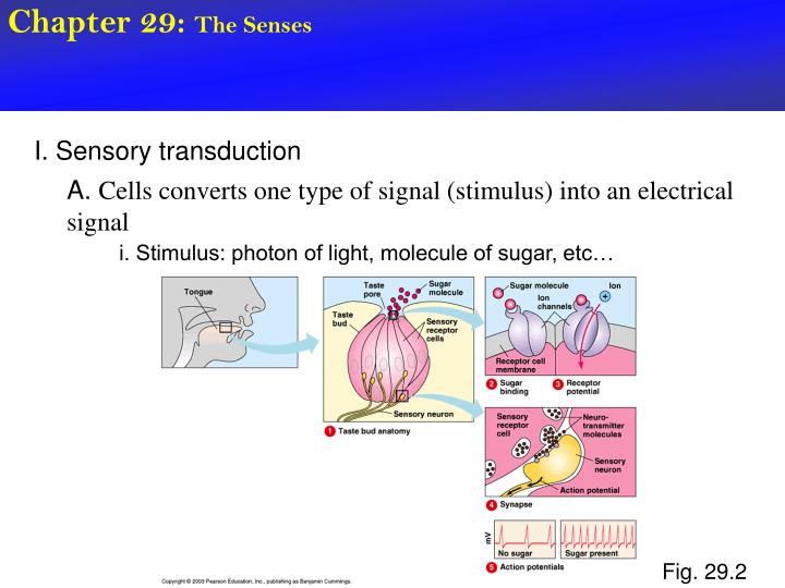
Chapter 29: The Senses. I. Sensory transduction. A. Cells converts one type of signal (stimulus) into an electrical signal. i. Stimulus: photon of light, molecule of sugar, etc…. Fig. 29.2. Chapter 29: The Senses. I. Sensory transduction.

E N D
Chapter 29: The Senses I. Sensory transduction A. Cells converts one type of signal (stimulus) into an electrical signal i. Stimulus: photon of light, molecule of sugar, etc… Fig. 29.2
Chapter 29: The Senses I. Sensory transduction A. Cell converts one type of signal (stimulus) into an electrical signal i. Stimulus: photon of light, molecule of sugar, etc… Fig. 29.2
Chapter 29: The Senses II. Sensory adaptation A. Tendency of a sensory receptor cell to become less sensitive to repeated stimulus i. Wear clothes without always being aware of it ii. “adjust” to a hot shower iii. “adjust” to smell in the room iv. Etc… B. Nervous system would become overloaded without it
Chapter 29: The Senses II. Five categories of stimuli A. Pain receptors B. Thermoreceptors C. Mechanoreceptors - touch, stretching, sound, pressure, motion, etc… - bend or stretch PM Fig. 29.3A
Chapter 29: The Senses II. Five categories of stimuli C. Mechanoreception by a “hair cell” - detect sound waves or movement of water - lateral line system - hearing and balance microvilli Fig. 29.3A
Chapter 29: The Senses II. Five categories of stimuli D. Chemoreceptors - nose/taste buds/ in arteries E. Electromagnetic receptors - electric currents in water - detect Earth’s magnetic field - photoreceptors - in eyes - detect photons
Chapter 29: The Senses III. Three types of eyes have evolved in invertebrates 1. Eye cup - simplest - found on planaria - detects only direction and intensity Fig. 29.4A
Chapter 29: The Senses III. Three types of eyes have evolved in invertebrates 2. Compound eye a. focuses light and forms images b. Ommatidia - tiny light detecting unit - each has own light focusing lens and photoreceptor cells - image formed in brain using combination of signals from all ommatidia Fig. 29.4B
Chapter 29: The Senses III. Three types of eyes have evolved in invertebrates 3. Single-lens eye a. Works like a camera b. pupil - small opening through which light passes c. iris - changes diameter of pupil (camera shutter) Squids, vertebrates - evolved independently Fig. 29.4B
Chapter 29: The Senses III. The vertebrate single lens eye 1. sclera i. Outer surface ii. Tough, whitish layer iii. Connective tissue 2. cornea i. Fuses to sclera at front of eye ii. clear iii. Lets light in, helps focus 3. choroid i. Pigmented layer below sclera ii. Forms iris at front of eye Fig. 29.5
Chapter 29: The Senses III. The vertebrate single lens eye Fig. 29.5 4. iris i. Gives eye its color ii. Regulate pupil size to adjust amount of light entering eye 5. lens i. Held in position by ligaments ii. Focuses images onto retina 6. Retina i. Layer below the choroid ii. Contains photoreceptor cells (it’s the film of the camera) - convert light to electric and send to optic nerve, which goes to brain iii. No photoreceptors where optic nerve attaches (blind spot) - two eyes, overlapping fields of view, fill in blind spot
Chapter 29: The Senses III. The vertebrate single lens eye Fig. 29.5 7. Two chambers i. Vitreous humor - fills large chamber behind lens - jellylike ii. Aqueous humor - small chamber in front of lens (b/w cornea and lens) - secreted by capillaries - brings oxygen, nutrients, etc… and removes wastes for cornea, lens, and iris cells. - glaucoma - increased pressure in eye caused by blockage of duct that drains aqueous humor iii. Humors help maintain shape
Chapter 29: The Senses III. The vertebrate single lens eye 8. Lacrimal gland i. Secrete tears - lubricate and clean the eye ii. Lacrimal sac drains tears into nasal cavity
Chapter 29: The Senses IV. Focusing light 1. mammals i. change the shape of the lens - thicker the lens the sharper light bends (muscles contract) 2. Fish and squid i. Change the physical position of the lens Fig. 29.6
Chapter 29: The Senses V. Artificial lenses or surgery can correct focusing problems 1. Visual acuity - read the eye chart from 20 feet away - from a distance of 20 feet, you can read the letters designated for 20 feet i. 20/20 - from a distance of 20 feet, you can read the letters that a person with 20/20 can only read from 10 feet ii. 20/10 iii. 20/50 - need to stand 20 feet to read letters that a person with 20/20 can read from 50 feets
Chapter 29: The Senses V. Artificial lenses or surgery can correct focusing problems 2. Most common visual problems i. nearsightedness, farsightedness, astigmatism - all are focusing problems - corrected with an artificial lens - named for type of vision that is UNIMPAIRED
Chapter 29: The Senses V. Artificial lenses or surgery can correct focusing problems 3. Nearsightedness (myopia) i. Eye is longer than normal - can’t flatten lens enough to see distant objects Fig. 29.7
Chapter 29: The Senses V. Artificial lenses or surgery can correct focusing problems 4. Farsighted (hyperopia) i. Eye is too short - can’t make lens thick enough to bend light onto retina Fig. 29.7
Chapter 29: The Senses V. Artificial lenses or surgery can correct focusing problems 5. Astigmatism i. Blurred vision caused by a misshapen lens or cornea Fig. 29.7
Chapter 29: The Senses VI. Photoreceptors of the eyes - rods and cones 1. Cones i. Stimulated by bright light (don’t function in night vision) ii. Distinguish color iii. 6 million cone cells per retina 2. rods i. Highly sensitive to light ii. Enable us to see in dim light (at night) iii. Shades of grey only iv. 125 million per retina Fig. 29.8
Chapter 29: The Senses VI. Photoreceptors of the eyes - rods and cones 3. Fovea i retina’s center of focus ii. Rods mostly on outer edge of retins iii. Cones mostly in center (fovea) - easier to see a star at night if you don’t look straight at it Fig. 29.8
Chapter 29: The Senses VI. Photoreceptors of the eyes - rods and cones 4. How do rods and cones detect light i. rhodopsin - visual pigment in discs of rod cells - can absorb dim light ii. photopsins - visual pigments in discs of cone cells - absorb bright, colored light Fig. 29.8 - There are 3 types of cone cells - each contains a different type of photopsin - blue cones, green cones, red cones - color blindness = deficiency in one or more of these types of cone cells.
Chapter 29: The Senses VI. Photoreceptors of the eyes - rods and cones 4. How do rods and cones detect light Fig. 29.8
Chapter 29: The Senses VII., Hearing and balance 1. Human ear a. Two separate organs i. hearing ii. balance iii. Both work by stimulating “hair cells” (microvilli) in fluid filled canals
Chapter 29: The Senses VII., Hearing and balance 1. Human ear b. Three regions i. Outer ear - pinna - auditory canal Fig. 29.9A
Chapter 29: The Senses VII., Hearing and balance 1. Human ear b. Three regions ii. Middle ear - eardrum Fig. 29.9B - separates outer ear from middle ear - sound waves vibrate ear drum, which vibrates the three bones - hammer, anvil and stirrup - oval window - membrane covered hole in skull - attached to stirrup - membrane vibrates when stirrup vibrates sending vibrations into the inner ear - Eustachian tube - conducts air b/w middle ear and back of throat - keeps pressure equal on both sides of ear drum
Chapter 29: The Senses VII., Hearing and balance 1. Human ear b. Three regions iii. Inner ear - fluid filled channels in bones of skull Fig. 29.9B - fluid set in motion by: 1. Sound waves (vibrating oval window) 2. Motion of the head
Chapter 29: The Senses VII., Hearing and balance 1. Human ear b. Three regions iii. Inner ear - cochlea (latin for snail) Fig. 29.9B - contains hearing organ (organ of Corti) - three fluid-filled canals Fig. 29.9C
Chapter 29: The Senses VII., Hearing and balance 1. Human ear c. Flow of sound Fig. 29.9B Vibrations in air -> collected by pinna and auditory canal -> vibrates ear drum -> hammer -> anvil ->stirrup -> oval window -> vibration of oval window produces pressure waves in fluid through upper canal to tip of cochlea and back through lower canal dissipating along the way. Fig. 29.9C
Chapter 29: The Senses VII., Hearing and balance 1. Human ear c. Flow of sound Fig. 29.9B - pressure wave through upper canal vibrates basilar membrane Result: hair cells brush back and forth on overlying membrane, bending microvilli and sending electrical signals Fig. 29.9C
Chapter 29: The Senses VII., Hearing and balance 1. Human ear d. Volume v. Pitch i. higher volume = higher amplitude of sound wave ii. higher pitch = higher frequency of sound wave
Chapter 29: The Senses VII., Hearing and balance 1. Human ear d. How does the ear determine volume? i. volume - high amplitude wave causes vigorous vibrations of fluid = high frequency of bending on microvilli - louder = higher frequency of signals sent to brain Fig. 29.9C
Chapter 29: The Senses VII., Hearing and balance 1. Human ear d. How does the ear determine volume? ii. Pitch Fig. 29.9B - basilar membrane is NOT uniform - varies from narrow and stiff to wide and flexible - different regions more sensitive to different pitches - brain determines pitch by which regions are sending most frequent signals Fig. 29.9C
Chapter 29: The Senses VII., Hearing and balance 1. Human ear e. Balance (position and movement) i. Semicircular canals ii. utricle iii. saccule Fig. 29.10 ALL filled with fluid
Chapter 29: The Senses VII., Hearing and balance 1. Human ear e. Balance (position and movement) i. Semicircular canals - three - detect changes in head rotation and angular movement Fig. 29.10 - arranged in three perpendicular planes - detect movement in all directions (X, Y, and Z) - hair cells located at base of each canal - microvilli projected into cupula (gelatinous mass) - as head moves, fluid moves, cupula moves, hair cells bend and signals are sent to brain.
Chapter 29: The Senses VII., Hearing and balance 1. Human ear e. Balance (position and movement) i. Utricle and saccule - detect position of head relative to gravity Fig. 29.10 utricle saccule
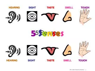
Chapter 29: The Senses VIII. Odor and taste 1. chemoreceptors a. One cell responds to a group of chemically related molecules (NOT JUST ONE) Fig. 29.12 Ex. One cell may detect 50 kinds of odors So how does the brain tell the difference b/w odors. - specific pattern of signals
Chapter 29: The Senses VIII. Odor and taste 2. Taste - Taste receptors in back of throat and taste buds on tongue - Types of taste receptors Sweet, sour, salty, bitter and umami (these detect amino acids) -Flavor interpreted by brain comes from a combination of signals from taste receptors
Chapter 29: The Senses VIII. Odor and taste 2. Taste - Taste receptors in back of throat and taste buds on tongue Fig. 29.12 - Types of taste receptors Sweet, sour, salty, bitter and umami (these detect amino acids) -Flavor interpreted by brain comes from a combination of signals from taste receptors INSECTS: taste with their feet - chemoreceptors in sensory hairs on their feet