Download
1 / 56
570 likes | 925 Views
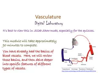
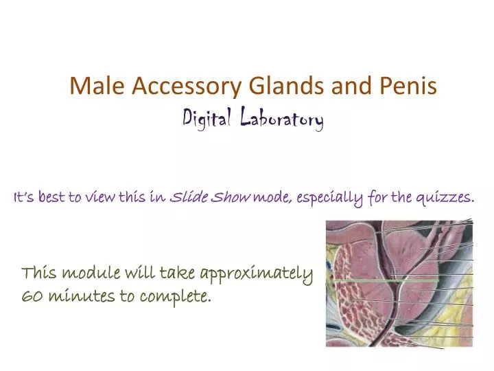
Male Accessory Glands and Penis Digital Laboratory. It’s best to view this in Slide Show mode, especially for the quizzes. This module will take approximately 60 minutes to complete. After completing this exercise, you should be able to:

E N D
Male Accessory Glands and Penis Digital Laboratory It’s best to view this in Slide Show mode, especially for the quizzes. This module will take approximately 60 minutes to complete.
After completing this exercise, you should be able to: • identify, at the light microscope level, each of the following: • Seminal vesicle • Mucosa • Muscularis • Adventitia • Prostate gland • Concretions • Urethral crest (colliculusseminalis) • Utricle • Ejaculatory ducts • Bulbourethral gland • skeletal muscle • mucous glands • Membranous urethra • Penis • Penile (spongy) urethra • Corpus spongiosum • Corpus cavernosum
MALE REPRODUCTIVE SYSTEM This is a reminder slide from the digital lab on testes and ducts. The urogenital diaphragm (indicated by the dotted green line) is a thin sheet of mostly skeletal muscle that includes the external urethral sphincter. Membranous refers to the part of the urethra that passes through the urogenital diaphragm. The membranous urethra is about 1cm in length. Major structures of the male reproductive system that allow for production and transmission of sperm include: Testis – produces spermatozoa Epididymis – storage and final maturation of spermatozoa Ductus (vas) deferens – transports spermatozoa to the prostatic urethra Urethra – has three parts in the male, prostatic, membranous, and penile, named for the structure that the urethra passes through
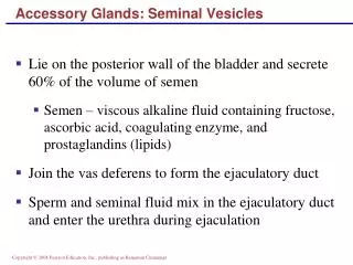
MALE REPRODUCTIVE SYSTEM This is a reminder slide from the digital lab on testes and ducts. These accessory glands and the penis will be covered in this module Accessory glands that are adjacent to the main pathway secrete fluids: Seminal vesicles – produce 65% of semen, its duct joins with the vas deference to become the ejaculatory duct, which passes through the prostate to drain into the prostatic urethra Prostate – produces 30% of semen, surrounds prostatic urethra Bulbourethral (Cowper’s) gland – secretes mucus during arousal for lubrication of the urethra
MALE REPRODUCTIVE SYSTEM – SEMINAL VESICLES adventitia mucosa muscularis mucosa adventitia muscularis adventitia Each seminal vesicle develops as an evagination from the vas deferens; each forming into a highly folded tubular structure. Sectioning the seminal vesicle reveals several apparently distinct lumina; however, note that these are all part of an interconnected lumen that becomes the duct of the seminal vesicle. The three regions of the wall of the seminal vesicle are (from inside to outside): Mucosa - epithelium plus loose connective tissue Muscularis – thick, fibrous smooth muscle layer that is very eosinophilic Adventitia – outer connective tissue Mucosa refers to an epithelium + underlying connective tissue that together form the inner lining of tubes that are inside the body but are exposed to the outside world; e.g. the inner lining of the respiratory and digestive tracts, and the urinary and reproductive structures all have a mucosa. Mucosa typically produces a secretion, so the surface of the mucosa is moist. Many of these organs also have a muscularis and adventitia as well.
MALE REPRODUCTIVE SYSTEM – SEMINAL VESICLES Mucosa muscularis These elaborate mucosal folds create numerous “pockets” that seem to be cut off from the main lumen (e.g. black arrows, there are many more in this image than indicated). Each of these is connected to the main lumen. Closer examination reveals that each region of the seminal vesicle contains numerous folds of mucosa. The folding of the mucosa in the seminal vesicles is often described as “lacey”. Each region is surrounded by brightly eosinophilic bundles of a fibrous smooth muscle – the muscularis. One of the borders between the muscularis and mucosa is indicated by the dotted line. Note that this is not the location of the basement membrane – the basement membrane is between the epithelium and connective tissue within the mucosa.
MALE REPRODUCTIVE SYSTEM – SEMINAL VESICLES muscularis Although the epithelium of the seminal vesicle is officially pseudostratified, the basal cells are less numerous, making the epithelium appear simple columnar. There is some variation in cell height, from cuboidal to columnar, but this is not as prominent as is the case for the efferent ductules. The epithelium is supported by loose connective tissue (arrows), which forms the core of the folds, and contains little, if any, smooth muscle. The muscularis that surrounds each region of the seminal vesicle is indicated.
MALE REPRODUCTIVE SYSTEM – SEMINAL VESICLES Video of seminal vesicle overview – SL56 Video of seminal vesicle – SL56 Video of seminal vesicle – SL161 • Link to SL 056and SL 161 • Be able to identify: • Seminal vesicle • Mucosa • Muscularis • Adventitia
MALE REPRODUCTIVE SYSTEM – PROSTATE GLAND The prostate gland has the same general features as the seminal vesicle; mucosa with pseudostratified epithelium, muscularis, adventitia. However, there are notable differences: The lumen contains noticable concretions (black arrows). The “lobules” of the prostate are much smaller, with noticeable smooth muscle interspersed between them. The folds of mucosa are much less elaborate. The epithelium undulates; the taller regions appear to be stratified (outlined), even though the epithelium, like the seminal vesicle, is pseudostratified
MALE REPRODUCTIVE SYSTEM – PROSTATE GLAND Video of prostate – SL56 Video of prostate – SL162 Video of child’s prostate – SL163 • Link to SL 056and SL 162and SL 163 • Be able to identify: • Prostate gland • Concretions • For the child’s prostate, just appreciate that the gland is not developed at this age. Specific recognition on a practical would be difficult.
MALE REPRODUCTIVE SYSTEM – PROSTATE GLAND To understand the orientation of SL162, it helps to get a more detailed understanding of the structures passing through the prostate. The area within the green rectangle is enlarged in the drawing on the right. Note that the short duct of each seminal vesicle (black arrow) joins with the vas (ductus) deferens to become the ejaculatory duct (blue arrow), which runs in the substance of the prostate gland to join with the prostatic urethra. Not shown is the prostatic utricle, a blind-ending remnant of the female reproductive tract (essentially the uterus) that is connected to the prostatic urethra at the same location as the ejaculatory ducts, but on the midline.
MALE REPRODUCTIVE SYSTEM – PROSTATE GLAND From Moore’s Anatomy text… These are posterior views with the rectum and other structures removed. Focus on the vas (ductus) deferens, seminal vesicles (glands), ejaculatory ducts, and prostatic utricle. In the lower image, note in particular that the posterior aspect of the prostate has been carved away to show the structures which run within the substance of the prostatic tissue.
MALE REPRODUCTIVE SYSTEM – PROSTATE GLAND Another Moore’s Anatomy image. The drawing to the right is similar to the drawing from two slides previous to this one, showing an ejaculatory duct passing through the prostate to join the prostatic urethra. The green line represents the section drawn below. Note that the prostatic urethra is “U-shaped”, due to the presence of a thickening of the posterior wall called the seminal colliculus. Our digital slide is a section similar to this, but taken slightly higher, so the prostatic utricle is near the prostatic urethra, but not joined with it. For this module, do not worry about the different zones of the prostate. The histological difference between them is subtle. However, these are extremely important clinically, and you should understand their structure and organization from your other presentations and references.
MALE REPRODUCTIVE SYSTEM – PROSTATE GLAND lumen lumen In our slide of the prostate gland in this region, the anterior wall is torn away, so you will be looking at the region inside the red rectangle. The urethral crest is outlined in red, the lumen of the prostatic urethra is indicated. Even at low power, you can see that the three structures indicated by the arrows have a more stratified-appearing epithelium than the surrounding glandular units. These structures are the utricle (green arrow) and ejaculatory ducts (black arrows).
MALE REPRODUCTIVE SYSTEM – PROSTATE GLAND Video of prostate ducts – SL162 • Link to SL 162 • Be able to identify: • Prostate gland • Concretions • Urethral crest (colliculusseminalis) • Utricle • Ejaculatory ducts
MALE REPRODUCTIVE SYSTEM – BULBOURETHRAL GLAND The urogenital diaphragm (indicated by the dotted green line) is a thin sheet of mostly skeletal muscle that includes the external urethral sphincter. The bulbourethral gland (Cowper’s gland) is a mucous gland that is embedded within the urogenital diaphragm. This combination, mucous gland plus skeletal muscle, is a feature that makes identifying this gland fairly easy.
MALE REPRODUCTIVE SYSTEM – BULBOURETHRAL GLAND As mentioned on the previous slide, the bulbourethral (Cowper’s) gland is pretty straightforward, because it contains mucous glands with interspersed skeletal muscle. Blood vessel (vein)
MALE REPRODUCTIVE SYSTEM – BULBOURETHRAL GLAND Video of bulbourethral gland – SL183 • Link to SL 183 • Be able to identify: • Bulbourethral (Cowper’s) gland • Mucus acini • Skeletal muscle
MALE REPRODUCTIVE SYSTEM – MEMBRANOUS URETHRA This is a reminder slide showing the membranous urethra. The urogenital diaphragm (indicated by the dotted green line) is a thin sheet of mostly skeletal muscle that includes the external urethral sphincter. Membranous refers to the part of the urethra that passes through the urogenital diaphragm. The membranous urethra is about 1cm in length.
MALE REPRODUCTIVE SYSTEM – MEMBRANOUS URETHRA V V • Like other organs that line internal spaces, the membranous urethra has three regions: • Mucosa (orange bracket), consisting of • an epithelium – here, the epithelium is stratified or pseudostratified, but varies considerably, so specific classification is difficult • Lamina propria – contains connective tissue and numerous venous sinuses (V) • Muscularis (green outlined areas) – smooth muscle • Adventitia – (black arrows) scant on most of this slide, will contain some skeletal muscle (area of blue arrow) that is part of the urogenital diaphragm / external urethral sphincter The urethra has mucous glands (of Littre), but these are not well-demonstrated on our slides.
MALE REPRODUCTIVE SYSTEM – MEMBRANOUS URETHRA Video of membranous urethra – SL164 • Link to SL 164 • Be able to identify: • Membranous urethra
MALE REPRODUCTIVE SYSTEM – PENIS The penis contains three erectile elements, two dorsal corpora cavernosaand a single ventral corpus spongiosum, that engorge with blood during erection. Each erectile element is composed of a dense connective tissue capsule (tunica albuginea) and venous sinuses. The penile (spongy) urethra passes through the corpus spongiosum.
MALE REPRODUCTIVE SYSTEM – PENIS V V U In these images, the corpora cavernosa (orange) and corpus spongiosum (green) are outlined. Like the membranous urethra, the penile (spongy) urethra (U) is lined by a stratified / pseudostratified epithelium. Each corpora contains connective tissue and numerous venous sinuses (V). The outer tunica albuginea of each is typically a dense irregular connective tissue, though you can clearly see scattered smooth muscle in the outer layer of the spongiosum (arrows). The corpora cavernosa tend to fuse toward the distal end of the penis, which is why it appears that these two structures are a single entity.
MALE REPRODUCTIVE SYSTEM – PENIS Video of penis – SL165 • Link to SL 165 • Be able to identify: • Penis • Corpus spongiosum • Penile (spongy) urethra • Corpora cavernosa
The next set of slides is a quiz for this module. You should review the structures covered in this module, and try to visualize each of these in light and electron micrographs. • Identify, at the light microscope level, each of the following: • Seminal vesicle • Mucosa • Muscularis • Adventitia • Prostate gland • Concretions • Urethral crest (colliculusseminalis) • Utricle • Ejaculatory ducts • Bulbourethral gland • skeletal muscle • mucous glands • Membranous urethra • Penis • Penile (spongy) urethra • Corpus spongiosum • Corpus cavernosum
Final quiz Self-check: Identify the organ. (advance slide for answers) Seminal vesicle
Final quiz Self-check: Identify the structure / organ. (advance slide for answers) Vas deferens
Final quiz Self-check: Identify the organ. (advance slide for answers) Prostate gland
Final quiz Self-check: Identify the cell indicated by the arrow. (advance slide for answers) Sertoli cell
Final quiz Self-check: Identify the structure indicated at X. (advance slide for answers) Prostatic utricle X
Final quiz Self-check: Identify the structures. (advance slide for answers) Efferent ductules
Final quiz Self-check: Identify the structure indicated by the X. (advance slide for answers) Penile (spongy) urethra X
Final quiz Self-check: Identify the cell indicated by the arrow. (advance slide for answers) spermatogonium
Final quiz Self-check: Identify the outlined structure. (advance slide for answers) Urethral crest X
Final quiz Self-check: Identify the organ. (advance slide for answers) Seminal vesicle
Final quiz Self-check: Identify the structure indicated by the X. (advance slide for answers) Corpus cavernosum X
Final quiz Self-check: Identify the structure indicated at X. (advance slide for answers) Ejaculatory duct X
Final quiz Self-check: Identify the organ. (advance slide for answers) Prostate gland
Final quiz Self-check: Identify the organ. (advance slide for answers) Bulbourethral (Cowper’s) gland
Final quiz Self-check: Identify the structure. (advance slide for answers) epididymis
Final quiz Self-check: Identify the organ. (advance slide for answers) Prostate gland
Final quiz Self-check: Identify the cell indicated by the arrow. (advance slide for answers) Leydig cells
Final quiz Self-check: Identify the outlined TISSUES. (advance slide for answers) Dense irregular connective tissue Smooth muscle
Final quiz Self-check: Identify the organ. (advance slide for answers) Bulbourethral (Cowper’s) gland
Final quiz Self-check: Identify the cell indicated by the arrow. (advance slide for answers) spermatid
Final quiz Self-check: Identify the structure / organ. (advance slide for answers) Vas deferens
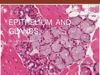
Final quiz Self-check: Identify the organ. (advance slide for answers) submandibular gland
Final quiz Self-check: Identify the organ. (advance slide for answers) Membranous urethra
Final quiz Self-check: Identify the cell indicated by the arrow. (advance slide for answers) Primary spermatocyte
Final quiz Self-check: Identify the outlined TISSUES. (advance slide for answers) Dense irregular connective tissue (elastic tissue) Skeletal muscle