Download
1 / 39
390 likes | 390 Views
Join us for a seminar on membrane functions presented by Dr. V. Valouskova from the Physiology Department at The 2nd Medical Faculty in Prague. Learn about diffusion rates, ion channels, passive and active transport, and more. Don't miss this opportunity to expand your knowledge in membrane physiology.

E N D
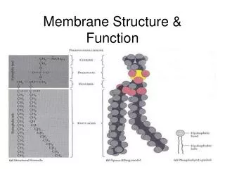
Membrane functionsseminar Dr. V. Valouskova Physiology Dept., The 2nd Medical Faculty, Prague
Diffusion rate J - final diffusion rate D - diffuse coefficient A - area Fick´s low (diffusion rate) J = -DA c/ x Stokes – Einstein equilibration D = kT/(6 r ) Dc/Dx - concentration gradient (difference in concentration /thickness of membrane minus - direction of diffusion k = Boltzman`s constant T = temperature r = molecule radius = viscosity Factors determining diffusion rate Diffusion surface (area) Concentration difference distance of diffusion size of molecules Viscosity (friction) temperature Lipid solubility Ion charge
fibrosis edema x η change A O2 O2 O2 CO2 x CO2 Examples of diffusion
Types of ion channels • Permanently opened- 2P potassium channels with 2 subunits (together with Cl- channels – resting MP) • Voltage gated – Na+,Ca2+, K+, Cl-, H+ channels- change conformation with voltage, they are opened (sometimes closed – e.g.. K+ channels in dendrites of neurons) by depolarisation, • Chemically gated – receptor-channel; ligand (receptor), mostly neurotransmitters: • receptors ionotropic- ion channel • receptor metabotropic – secondary messenger systems (G-proteins, IP3, etc.) • pHgated (pain) • mechanically gated • gated by other forms of energy– e.g.. thermal energy Paramoecium (protozoa) bangs into smt Ca+2 channels are mechanically opened - membrane depolarisation – cilia - backward movement pushes forward K+ - hyperpolarisation – speeds up forward movement
K+channels opened (2 subunits) - 2P-K channels – resting MP inward rectifiers(- KIR) - one direction permeable voltage-modulated (Kv) (4 subunits) voltage-dependent- 2 main levels : closed – opened gated – conformation changes K+ channel closedopened
Na+ channels Voltage gated Inactivated 2. Activated = opened 1. Resting = closed Epithelial Na channel (ENaC) – three subunits a, b, g. a- transfer of Na+, b a g assist to subunit a increase transport of Na+ .(i.g. in kidney - regulate ion of ECT volume, aldosteron)
Ionotropic ligand-gated channels Ligand opens ion channel directly i.e. acetylcholine receptor- nicotine type - channel opens when 2 molecules of ACh were bound to receptors – permeability for ionts
Metabotropní kanály Metabotropic ligand-gated channel Ligand opens ion channel indirectly i.e. during signal reception and signal (information) processing and storage
Passive - active transport facilitated diffusion [xy]1 > [xy]2 [xy]1 [xy]2 primary secondary
Proton pump Difference in electrochemic potencials of protons H+(secondary of other ions) is, besides ATP, universal source of energy which cells use
Secondary active transport(symport, antiport) - examples Symport Na+/glukose1:1 kidney, intestine Activation – Na+ Na, K gradient -> used for transport Antiport Na+/Ca2+ 3:1 Heart Activates- kalmodulin active Antiport Na+/H+ Each cell– cell membrane Activation – level of H+ in cytosol symport sacharides, amino acids Antiport Cl - / HCO3- Erytrocyt Activation – level of HCO3- in erytrocyte
1. Pancreas Langerhans cells - b cell K+channels Ca2+channels -70 mV GLUT K+channels
2. Pancreas - b cell K+channels Ca2+channels -70 mV GLUT GLUCOSE K+channels
3. Pancreas - b cell K+channels ATP closes channel Ca2+channels ATP -70 mV GLUT GLUCOSE K+channels
4. Pancreas - b cell depolarization depolarization K+channels - - - - - - - - - Ca2+channels opened + + + + + + ATP GLUT GLUCOSE K+channels
5. Ca2+ [Ca2+ ]IC Pancreas - b cell depolarization depolarization K+channels - - - - - - - - - Ca2+channels opened + + + + + + ATP GLUT GLUCOSE K+channels
6. Ca2+ [Ca2+ ]IC K+channels Pancreas - b cell depolarization K+channels - - - - - - - - - Ca2+channels opened + + + + + + ATP GLUT GLUCOSE insulin
7. Pancreas - b cell depolarization Ca2+ K+channels - - - - - - - - - Ca2+channels + + + + + + ATP [Ca2+ ]IC GLUT GLUCOSE K+channels (Ca+2 dependent ) insulin repolarization ( Ca +2 channels closed)
Pancreas Langerhans cells - b cell 8. depolarization Ca2+ K+channels - - - - - - - - - ATP dependent Ca2+channels + + + + + + voltage dependent ATP [Ca2+ ]IC GLUT GLUCOSE facilitated diffusion insulin K+channels Ca+2 dependent exocytose repolarization ( Ca +2 channels closed)
Proximal tubule Na+/K+ ATPase!!! 3 Na+ ATP 2 K+ PROXIMAL TUBULE lumen blood Primary urine Epithelial cell Kidney ? [Na+]in ?
antiport antiport symport Na++ Glu, AMK (cations) primaryactive transport - LNTP + secondaryactive transport PROXIMAL TUBULE lumen blood H+ elimination Na+/K+ ATPase !!! Na+/H+ transport (electroneutral) ATP LNTP -lumen negative transport potential Na+ resorption Active or passive transport ??
Na++ Glu, AMK (cations) PROXIMAL TUBULE lumen blood H+ elimination Na+/K+ ATPase !!! Na+/H+ exchange (electroneutral) ATP Na+ resorption - LNTP + Cl-resorption Cl- paracellular pathway; followed by H2O
Na++ Glu, AMK (cations) PROXIMAL TUBULE lumen blood H+ elimination Na+/K+ ATPase !!! Na+/H+ exchange (electroneutral) ATP Na+ resorption - LNTP + Cl-resorption Cl- paracellular pathway; followed by H2O + LPTP - LPTP -lumen positive transport potential
PROXIMAL TUBULE lumen blood H+ elimination Na+/K+ ATPase !!! Na+/H+ exchange (electroneutral) antiport antiport ATP Na++ Glu, AMK symport Na+ resorption - LNTP + Cl-resorption Cl- paracellular pathway; followed by H2O simple diffusion + LPTP - ?? Ca2+,Mg2+,Na+,K+ resorption Ca2+, Mg2+, Na+, K+ paracellular, followed by H2O
parathormon secondaryactive transport primaryactive transport PROXIMAL TUBULE lumen blood H+ elimination Na+/K+ ATPase !!! Na+/H+ exchange (electroneutral) antiport antiport ATP symport Na++ Glu, AMK Na+ resorption - LNTP + Cl-resorption Cl- paracellular pathway; followed by H2O simple diffusion + LPTP - Ca2+,Mg2+,Na+,K+ resorption Ca2+, Mg2+, Na+, K+ paracellular, followed by H2O PO43-resorption Na+/K+ ATPase Na++ Pi cotransport (secondary AT) ATP
Resting membrane potential = Electro-chemicequilibration mi [xi]II ~ mi = RT ln + nFD [xi]I Two parts - diffusion (osmotic) work, electric work, replacement of some amount of electric charges between 2 solutions Electro-chemical potential for the ion [xi] – ion concentration xi in solutions I a II, F - Faraday´s constant , n (or z) quantivalence of ion (e.g.. n=+1 for K+and –1 for Cl-). electric potential in Volts, i.e. MEMBRANE POTENTIAL.
Origin of resting MP Membrane = demarcation line between two different environments – shift of electrically charged chemic elements - ions Membrane – resting stage - only K+ ions permeability (permeability for Cl- and Na+ non-significant) K+ ions tendency to diffuse to the place with lower concentration complementary A-(protein anions) cannot accompany K+ Potential – (charge difference) slows down K+ ions outward (Coulomb´s low) - inside - negative outside positive Osmotic power (chemic gradient) forces K+ outward (down the concentration gradient) reverse power (electric) – forces K+inward dynamic equilibration = equalizing electric and chemic gradient (electro-chemic equilibration) http://www2.biomed.cas.cz/d331/vade/
Nernst´s equilibration ECl = equilibration potential for Cl– R = gas constant T = absolute temperature F = Faraday`s constant(number of coulombs per mol) ZCl = quantivalence Cl– (–1) [Clo–] = concentration Cl– out, [Cli–] = concentration Cl– inside of cell
Goldman´s equilibration membrane potential V - membrane potential, R – gas constant, T - absolute temperature, F - Faraday constant PK+, PNa+ a PCl– permeability for K+, Na+ a Cl–, respectively [ ] concentration index i-in and o-out (intra-, extracellular concentration)
Hyperkalemia K+ + K+ K+ - more than 5 (10) minutes chemic gradient elektric gradient Plasma hyperkalemia resulted in membrane depolarisation. Underlying process?? Normokalemia MP - 90mV RMP - 70mV RMP - 80mV Hyperkalemia Proteins- Proteins- Proteins- K+ - - - K+ K+ K+ K+ K+ K+ K+ K+ - - - - - - + + - - - - - - - - - - New equilibrium between electric and chemic driving forces is established Membrane is DEPOLARISED - seconds to 5 (10) minutes Gradual (long-lasting) increase of [K+]e Quick experimental increase of [K+]e
Electric impulses in the NS • LOCAL POTENTIALS OR CURRENTS - graded, spreading with decrement • generator or receptor potentials - sensory terminals – transduction of energy i.e. mechanic or thermal to electric (graded according to the number of activated receptor cells and intensity of activation);foto-, chemo-, mechano-transduction • (post)synaptic potential (current), graded according to • - number ofexcreted quanta of neuromediators: • inhibitory (hyperpolarisation of postsynaptic membrane several ms - Cl channels) • excitatory (depolarisation – Na and/or Ca channels) • - number of active receptors (postsynaptic membrane) • 2. ACTION POTENTIALS (spikes)
Synaptic potential – sum of „channels“ potentials Haines, Fundamentel Neuroscience, 1997, p. 45
Ion membrane permeability during AP(initial segment) Action potential Na+ current inward K+ current outward permeability 1 – subthreshold depolarisation (EPSP) – K+>Na+ 2 – threshold depol. EPSP-peak – Na+ permeability = K+ permeability (open arrow) gNa g K - permeability for Na, K ions, respectively 3 – action potential (AP) 4 – subthreshold depolarisation (EPSP) – shorter latency Closed arrow –peak of AP - permeability K+ = Na+ Haines, Fundamental Neuroscience, 1997, p. 37
Refractory period Haines, Fundamentel Neuroscience, 1997, p. 38
Spontaneous neuron activity– intracellular registration out -70 mV in