Download
1 / 55
550 likes | 715 Views
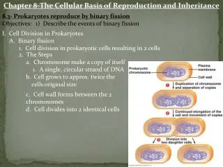
Chapter 8. The cellular basis of reproduction and inheritance. Cell Division. Ratio of surface area to volume cannot get too large because… Demands on DNA = “info crisis” Nutrient and waste movement = “traffic jams”. When demands are too high The Result: Cell Division.

E N D
Chapter 8 The cellular basis of reproduction and inheritance
Cell Division • Ratio of surface area to volume cannot get too large because… • Demands on DNA = “info crisis” • Nutrient and waste movement = “traffic jams” When demands are too high The Result: Cell Division
Cell Division in Our Bodies • Brain cell - doesn’t divide in a lifetime • RBC’s - 120 days • Skin cells - 35 days • Stomach cell - 2 days • Bacteria cell - 20 to 30 minutes
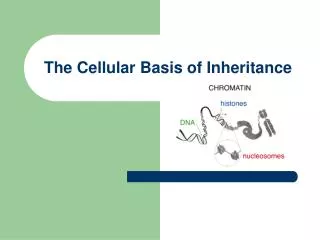
DNA PackagingSimple to complex • Double helix • Chromatin: DNA and histones (protein) • Supercoils • Chromosomes: tightest packaged DNA
Chromosomes Genetic information in eukaryotic cells is organized into chromosomes. Chromosomes are only visible (with a light microscope) during cell division, it is then that they pack tightly into a dense visible structure. Chromosomes are copied before division Copied chromosomes consist of two identical “sister” chromatids held together by an area called the centromere.
Parts of a Homologous Pair of Chromosomes Chromosome Centromere Chromatid Homologous Chromosomes
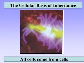
Cell Division • “All cells come from pre-existing cells” • Prokaryotic cell division: binary fission • Eukaryotic cell division: mitosis and meiosis
Cell Division • Why do cells divide? • To repair damaged or old cells • So the organism can get larger (grow)
The Cell Cycle Describes the Life Cycle of a Cell • Every cell follows a specific sequence of steps, just like a human goes through stages of life.
The Major and Minor Stages of the Cell Cycle • Interphase:Cell growth and DNA replication • Gap 1 • Synthesis • Gap 2 • The Mitotic Phase: • Mitosis:the division of the nucleus • Prophase • Metaphase • Anaphase • Telophase • Cytokinesis: the division of the cytoplasm.
Interphase(Stage 1) 3 Sub-Stages of Interphase • Gap 1 (G1) – Cell grows, preps for DNA replication • Synthesis phase (S-phase) – DNA is replicated • Gap 2 (G2) – Cell grows, preps for mitosis
Mitosis (Stage 2 – part of the mitotic phase) • Prophase • Metaphase • Anaphase • Telophase • PMAT! 4 Sub-Stages of Mitosis
Mitosis: The division of the nucleus • Prophase • Chromosomes become visible • Centriolesseparate and move to opposite ends of the cell • The spindle(a structure that helps to separate the chromosomes) forms • Nucleolus disappears and nuclear envelope breaks down • Metaphase • Microtubles attach to the centromeresto move the chromosomes • Chromosomes line up in the center of the cell
Mitosis: The division of the nucleus • Anaphase • Centromeresthat join sister chromatids split in half • Sister chromatids split and become individual chromosomes • Chromosomes move to opposite poles of the cell • Telophase • Chromosomes begin to disperse and are less visible • Nuclear envelopes begin to reform around each cluster of chromosomes • The spindle fibers dissolve
At the end of Interphase Mitosis Telophase Metaphase Anaphase Prophase At the end of Interphase Metaphase Prophase Anaphase Telophase Blood lilly Mitosis
Cytokinesis(Stage 2 – part of the mitotic phase) • The result of Mitosis is the formation of two nuclei each with duplicate sets of chromosomes formed within the cytoplasm of a single cell. • Cytokinesisseparates the cytoplasm of the cell into two. • During cytokinesis in plant cell a cell plate forms between the nuclei • During cytokinesis in animals a cleavage furrow forms pinching the two cells apart
Do You Know the Stages of Mitosis? CYTOKINESIS TELOPHASE PROPHASE ANAPHASE INTERPHASE ANAPHASE METAPHASE
Controls on cell division • Anchorage dependence – cells must be in contact with a solid surface to divide. • Density dependent inhibition – when cells are touching other cells they don’t usually divide • Growth factors are proteins that tell cells to divide
Growth factors control the cell cycle • Cells do not usually divide unless they get a chemical signal. • The cell cycle control system is a set of molecules that help to trigger and coordinate important steps in the cell cycle. • Checkpoints are critical control points in the cycle when cells will either stop or go forward in the cycle. • Proteins that control the steps in the cell cycle are called cyclins.
Controls on Cell Division • Most cells are in interphase • Checkpoints for division: • Is DNA fully replicated? • Is the DNA damaged? • Are there enough nutrients to support cell growth • Proto-oncogenes – start cell division • Tumor-SuppressorGenes– stop cell division Mutations in both types of genes can lead to cancer
Controls on Cell Division • Example Tumor suppressor gene: • p53 stops cell division and induces apoptosis in abnormal cells (cells with damaged DNA) • Apoptosis – cell suicide (pre-programmed death) • Example proto-oncogene: • Ki-ras – when mutated can lead to increased risk of lung, ovarian, colon and pancreatic cancer Apoptosis Video
Uncontrolled Cell Growth • Cancer is a disease caused by cells that do not respond to the signals that regulate growth. • According to the American Cancer Society: • As of 2008 in the USA a man has a 1 in 2 chance of developing cancer in his lifetime and a woman has a 1 in 3 chance of developing cancer in her lifetime • Cancer accounts for 1 in 4 deaths in the U.S. each year.
Cancer occurs when extra cells form a mass or a tumor • Benign tumor remain within the mass (non-cancerous) • Malignant tumor cells invade and destroy healthy tissues elsewhere in the body (cancerous)
Types of Cancer Normal Cells • Carcinoma-affects skin cells (most common) • Sarcoma- affects muscle, bone, cartilage, fat or connective tissues. • Leukemia- affects white blood cells or their precursors (begins in bone marrow) • Lymphoma- affects bone marrow cells/lymphatic system (solid tumors) Metastasized Tumor Malignant Tumor
What Causes Cancer? • Gene changes due to: • diet – lack of antioxidants • tobacco use • exposure to radiation • exposure to chemicals • Inherited genes (p53 or Ki-ras don’t work) • Immune System Failure Carcinogens
Treatment Side Effects Surgery: physically remove tumor Restricted to a few types of cancer, may not get all cells, damages organs. Radiation Therapy: Damages cancer cell’s DNA, cells won’t reproduce Can injure or kill healthy cells Chemotherapy: Chemicals target cancer cells given 2 or more at a time Similar to radiation therapy Hormone Therapy: Blocks hormones that cancer needs to grow Weakening of the bones can also effect sex characteristics Biological Therapy: Elicits a response from your immune system (helps immune system fight cancerous cells) Allergic reactions, swelling, itching also flu like symptoms, greatest concern is extreme allergic reactions.
Cancer Among Men • The three most common cancers among men include: • Prostate cancer • Lung cancer • Colorectal cancer • The leading causes of cancer death among men are: • Lung cancer • Prostate cancer • Liver cancer
Cancer Among Women • The three most common cancers among women include: • Breast cancer • Lung cancer • Colorectal cancer • The leading causes of cancer death among women are: • Lung cancer • Breast cancer • Colorectal cancer
Meiosis and Crossing Over • Mitosis is cell division to replace dead cells or allow an organism to grow. • Meiosis as a specialized type of cell division that occurs only in the reproductive organs, to create reproductive cells.
Chromosomes in Somatic Cells are Matched in Homologous Pairs • Somatic cell: a body cell • Ex. skin cell, bone cell, etc. • Contain 46 chromosomes (diploid meaning 2 copies) • Pairs 1-22 are called autosomes • Pair 23 (X and Y) are sex chromosomes • There are 23 types of chromosomes which exist as pairs called homologuschromosomes
Homologous chromosomes • Carry the same genes at the same locations (called loci) • Genes may be different versions • Ex. gene is eye color versions of those genes include: brown, blue, grey, hazel, green
Gametes have a single set of chromosomes • Gametes are sex cells • Sperm cell and egg cell • Gametes have only 1 copy of each type of chromosome (called haploid – 1n) • When two gamete cells combine a diploid cell (2n) called a zygote is created.
Meiosis reduces the chromosome number from diploid to haploid • Meiosis • Occurs only in sex cells • Two divisions • Meiosis I • Meiosis II • Creates haploid cells from diploid cells • Creates genetic diversity through: • Crossing over (Prophase I) • Independent assortment (Metaphase I)
STEPS OF MEIOSISMEIOSIS I PROPHASE I METAPHASE I ANAPHASE I TELOPHASE I
Tetrad STEPS OF MEIOSIS: MEIOSIS I PROPHASE I: Grouping of tetrads and crossing over Paternal Chromosome Maternal Chromosome Homologous chromosomes sister chromatids sister chromatids
Non-sister chromatids Crossingover STEPS OF MEIOSIS: MEIOSIS I PROPHASE I: Grouping of tetrads and crossing over Crossing over: homologous chromosomes exchange genetic material
Crossing over contributes to genetic diversity spindle fiber centrioles STEPS OF MEIOSIS: MEIOSIS I PROPHASE I: Grouping of tetrads and crossing over
OR STEPS OF MEIOSIS: MEIOSIS I METAPHASE I: Independent assortment • Independent Assortment: tetrads line up randomly during Metaphase I • Contributes to genetic diversity
STEPS OF MEIOSIS: MEIOSIS I ANAPHASE I: Homologous chromosomes separate • Tetrads split • Homologous chromosomes move to opposite ends of cell • Sister chromatids stay together (not like mitosis)
STEPS OF MEIOSIS: MEIOSIS I TELOPHASE I/CYTOKINESIS I: Homologous chromosomes separate • 2 new cells each with haploid set of chromosomes • In human sex cells 23 chromosomes
MEIOSIS I – recap! PROPHASE I METAPHASE I ANAPHASE I TELOPHASE I
MEIOSIS I – recap! Q: What is the purpose of Meiosis I? A: To decrease the # of chromosomes by 1/2 Q: What 2 features of Meiosis I increase the diversity of the daughter cells? A: Crossing Over & Independent Assortment
PROPHASE II METAPHASE II ANAPHASE II TELOPHASE II MEIOSIS II
STEPS OF MEIOSIS: MEIOSIS II • Happens immediately after cytokinesis I – interphase does not happen! • Meiosis II stages: • prophase II • metaphase II • anaphase II • telophase II • Meiosis II is nearly identical to Mitosis
MEIOSIS II PROPHASE II METAPHASE II ANAPHASE II TELOPHASE II
Summary of MEIOSIS The entire process of meiosis results in four haploid daughter cells. gametes = sperm or egg
Karyotypes • A karyotypeis a photographic inventory of an individual’s chromosomes • White blood cells are made to go through mitosis. Cells are treated to stop cells during metaphase of mitosis and a picture is taken of the cell. • Karyotypes are used to determine gender and also identify chromosomal abnormalities.