Download
1 / 20
200 likes | 406 Views
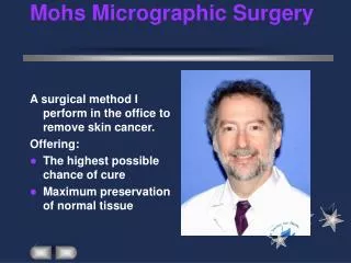
MOHS MICROGRAPHIC SURGERY (MMS). Hayleigh Gordon Histology BMS 1. What is MOH’s ?. Mohs surgery or MMS is a specialised technique enabling the surgeon to remove a skin tumour in several stages. Without removing too much healthy tissue.

E N D
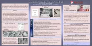
MOHS MICROGRAPHIC SURGERY (MMS) Hayleigh Gordon Histology BMS 1
What is MOH’s ? • Mohs surgery or MMS is a specialised technique enabling the surgeon to remove a skin tumour in several stages. Without removing too much healthy tissue. • Technique was pioneered by Fredrick Moh, an American Surgeon in the late 1940’s. • It is called micrographic surgery as each piece of skin removed is checked and examines using a microscope.
All Kent MOH skin procedures take place at Canterbury Hospital, this is a very specialist surgery and is only available in a few hospitals within the UK. • There are two surgery procedure rooms, both with quick access to the moh room
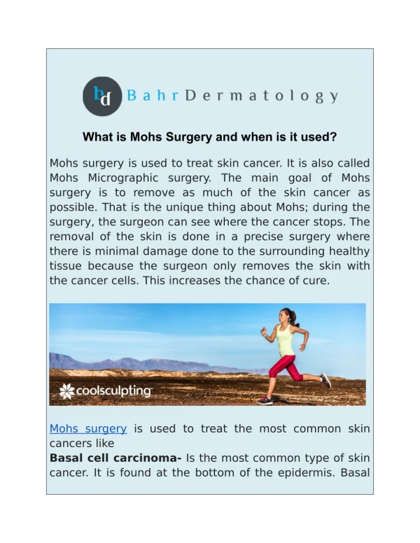
WHY USE MOHS? • Mohs micrographic surgery is commonly used to remove large tumours, tumours in hard to treat places as well as recurrent tumours. • It is commonly used on Basel Cell Carcinomas (BCC), it can be also be used on Squamous Cell Carcinomas (SCC) as well as Melanomas in some cases.
What is a BCC? • Basal cell Carcinomas are the most common cancer found in humans. • It arises from the basal cell layer of the skin, in the majority of cases it grows slowly. • Fortunately these tumours virtually never spread internally or present life threatening. They can however grow to be unsightly, or cause facial distortion. • Most cases of BCC are related to exposure to sunlight or repetitive sunburn injury. There are however a few genetic disorders such as Gorlin’s syndrome which lead to a tendency to develop BCC’s.
Mohs surgery cont… • The removed tissue is carefully mapped on a schematic drawing. • The tissue is colour coded using different tissue marking paints to identify the different margins.
Moh surgery cont… • Once inked the tissue is flattened as much as possible to ensure the whole tissue margins can be cut and seen when stained. • A chuck is covered in OCT, a medium to hold the tissue securely. The tissue is added onto the chuck an placed into a widget. The widget is linked to a liquid nitrogen canister, which is controlled by a foot pedal. Whilst pressing this, the surgeon flattens the tissue. The liquid nitrogen freezes the surrounding medium and the tissue, holding it securely in place.
Equipment Liquid nitrogen canister Widget Foot pedal attachment
Mohs procedure • The frozen chuck is then given to the Moh assistant to be placed into the Cryostat. • Tissue is cut quickly and efficiently. • The surgeon may perform 3- 4 patient procedures in short succession, the assistant must keep the chucks in order with clear patient identification.
Cryostat Temperature monitor External wheel for movement, enabling Efficient cutting of a section Tools (kept cold) Storage for chucks Chuck holder Blade Controls thickness Of sections in micrometres .
Cryostat • A cryostat is a microtome within a freezer, sections are cut at a certain temperate usually between -20 and -30 Celsius. • Once frozen, the specimen on the chuck is mounted on the microtome. The external wheel is rotated and the specimen advances toward the cutting blade. Once the specimen is cut to a show ‘full face’ therefore the whole section can be seen including a full epidermis, it is mounted on a warm (room temperature) clear glass slide, where it will instantaneously melt and adhere. These can then be placed onto the linear strainer.
Staining • A Haematoxylin and Eosin stain is used to aid medical diagnosis. Haematoxylin stains nuclei of cells blue, eosin is the counter stain which stains the other structures various shades of red or pink. • As the sections are cut they are placed on a liner stainer, this is quicker than a routine use laboratory stainer. It takes 10-15 minutes to travel through the required solutions.
Liner Stainer Bicycle chain to attach slide holder Eosin Haematoxylin Alcohol
Mohs procedure cont… Stained section on slides Microscopic View
Mohs procedure • Once the slides have been efficiently stained they are held within a xylene bath. The slides are then removed and quickly cover slipped. They are placed within slide trays in order. • They are placed by the microscope for rapid diagnosis. The surgeon thoroughly examines the slide microscopically. 100% of the tissue margin is examined and evaluated, to ensure whole tumour removal. If cancer cells are seen the area is marked on the schematic drawing and the process is repeated after removal of more tissue from the patient.
Microscope Lens Main microscope view Second lens Slide camera objective Slide platform
Moh Benefits • The process is designed to be quick for rapid and effective removal. The surgeon will remove skin until they are confident there is no tumour left behind. • For certain skin cancers Mohs procedures have a cure rate of up to 99%. It is precise while sparing of healthy tissue. Therefore scarring is kept minimal also. • The procedure enables you to go home the same day as your operation, providing everything goes to plan
Moh Risks • As with any surgery there is always risk. However with the lowest recurrence rate it is seen to be extremely beneficial. • The procedure can be lengthy and is performed under local anaesthetic which carries its own risks to each patient.