Download
1 / 13
150 likes | 525 Views
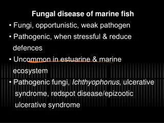
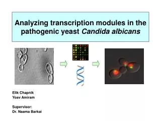
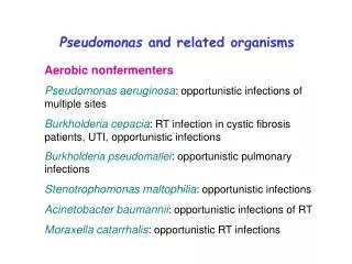
OPPOTUNISTIC MYOCOSIS Pathogenic yeast (opportunistic) I. Candidiasis - Moniliasis Etiologic agent: 1. Candida species. 2. candida albicans The most common causative agent. Morphology: Yeat like cells - reproduce by budding, oval cells. Diseases. . 1. Oral Candidiasis – mouth.

E N D
OPPOTUNISTIC MYOCOSIS Pathogenic yeast (opportunistic) I. Candidiasis - Moniliasis Etiologic agent: 1. Candida species. • 2. candidaalbicans The most common causative agent. Morphology: Yeat like cells - reproduce by budding, oval cells. Diseases.
1. Oral Candidiasis – mouth Infant – newborn Older children and adult • Thrush appear as creamywhite patches or grey membrane covering tongue • Acquired through passage in infected vagina. Thruth Predisposing Factors
2.Vaginitis: vulvovaginitis Similar to thrush: yellow milky vaginal discharge with irritation and itching. Predisposing factors: • Diabetes • Antibiotics • Pregnancy • Oral contraceptic
3. AlimontaryCandiasis • Usually follow antibiotic administration. • Or any predisposing factor listed for oral candiasis. • Inflamation of the perianal region (region around the anus) intecfioñmay.extend to buttocks and thighs. 4. Cutaneous and Systemic candiasis: Skin lesion usually occur in worm, moist folds of skin (axilla, groin) such as in common in fat people and diabetics.
Nail infection: • Painful, reddened swelling lesion. Nail becomes thick and brittle. 5. Systemic Candiasis: Common in patients receiving immunosupressive drugs (diabetes) • Urinary tract infectian. Also mechaniclly by catheterization. • Endocarditis: Also mechanically by implanted prosthetic devices (valves). • Kidney infection and Meningitis.
Diagnosis: • Sputum, Skin scrapping, swabs. • Do gram stain – look for gm positive budding oval cells and pseudohyphae. • Skin scrapping treated with 10 KOH. Culture: blood agar, sabourauds' agar at 37οC and RT. Candida albicans can be distinguished from other candida species by the formation of germtubes when cultivated in serum at 37°C for 2 - 21/2 hours. Examine microscopically.
Treatment: • Oral thrush can be treated by Nystatin - Orally Systemic infection: Amphotericin B in combination with flucytosine. • Miconazole, ketoconazole orally. • Oral Infection give multivitamins in particular B2.
II.Cyptococcossis: Atiologic agent: Cryptoccusneoformans, a capsulated yeast saprophytic in soil. Epidemiology and pathogenesis: infection acquired by the inhalation of the fungus in dried pigeon feces. More commonly in IMMUNOSUPRESSED PEOPLE.
Lung Pneumonia Spread to blood stream
Diagnosis: • India ink wet – preparation – look for capsulated yeast. The presence of this in sedimented C.S.F. Considered as a presumptive diagnosis. • Cultvation of the organism in sabouraud or blood agar. • Latex agglutination: Latex coated with antigen can be agglutinated by antisera.
Treatment: • Combination of amphotericuin B and 5 – Fluorocytosine untreated meningitis may be fatal.
II. Zygomycosis (older name mucormycosis, phycomycosis) Human infection usually caused by: Rhizopus, and Mucor. • Infection acquired by inhalation of spores. • Produce OPPORTUNISTIC FUNGI (mold) Fetal Meningits Pulmonary infection Blood & other internal organs
Diagnosis: Organism in tissue detected by staining with hematoxyllin - eosin observe for non septatehyphae. Treatment: Very fatal due to rapid spreading. • Amphotericin B. • Surgery.