Download
1 / 1
10 likes | 132 Views
NIRT: Active Nanoparticles in Nanostructured Materials Enabling Advances in Renewable Energy and Environmental Remediation. David A. Dixon, James L. Gole, Andrei Federov, Clemens Burda, Greg Szulczewski

E N D
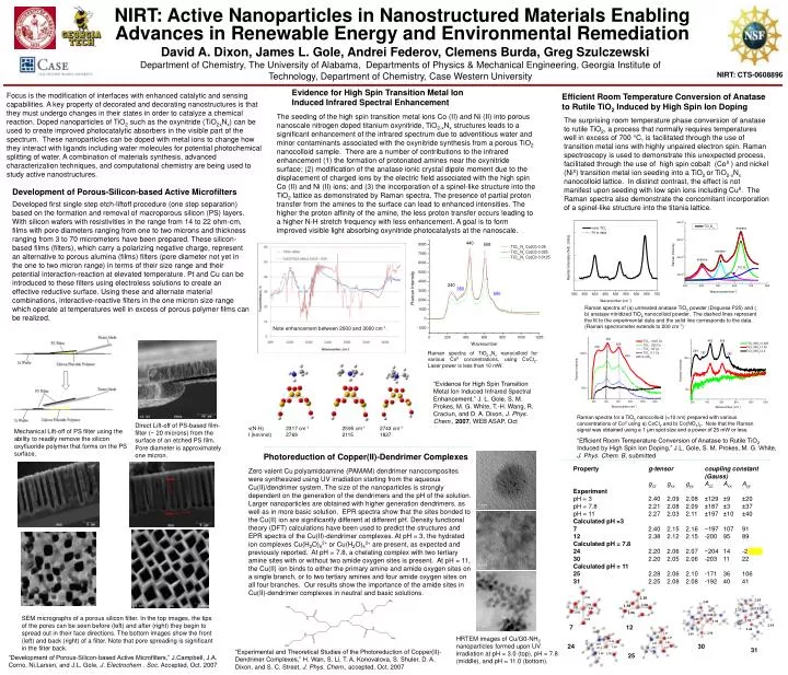
NIRT: Active Nanoparticles in Nanostructured Materials Enabling Advances in Renewable Energy and Environmental Remediation • David A. Dixon, James L. Gole, Andrei Federov, Clemens Burda, Greg Szulczewski • Department of Chemistry, The University of Alabama, Departments of Physics & Mechanical Engineering, Georgia Institute of Technology, Department of Chemistry, Case Western University NIRT: CTS-0608896 Evidence for High Spin Transition Metal Ion Induced Infrared Spectral Enhancement Focus is the modification of interfaces with enhanced catalytic and sensing capabilities. A key property of decorated and decorating nanostructures is that they must undergo changes in their states in order to catalyze a chemical reaction. Doped nanoparticles of TiO2 such as the oxynitride (TiO2xNx) can be used to create improved photocatalytic absorbers in the visible part of the spectrum. These nanoparticles can be doped with metal ions to change how they interact with ligands including water molecules for potential photochemical splitting of water. A combination of materials synthesis, advanced characterization techniques, and computational chemistry are being used to study active nanostructures. Efficient Room Temperature Conversion of Anatase to Rutile TiO2 Induced by High Spin Ion Doping The seeding of the high spin transition metal ions Co (II) and Ni (II) into porous nanoscale nitrogen doped titanium oxynitride, TiO2-xNx structures leads to a significant enhancement of the infrared spectrum due to adventitious water and minor contaminants associated with the oxynitride synthesis from a porous TiO2 nanocolloid sample. There are a number of contributions to the infrared enhancement (1) the formation of protonated amines near the oxynitride surface; (2) modification of the anatase ionic crystal dipole moment due to the displacement of charged ions by the electric field associated with the high spin Co (II) and Ni (II) ions; and (3) the incorporation of a spinel-like structure into the TiO2 lattice as demonstrated by Raman spectra. The presence of partial proton transfer from the amines to the surface can lead to enhanced intensities. The higher the proton affinity of the amine, the less proton transfer occurs leading to a higher N-H stretch frequency with less enhancement. A goal is to form improved visible light absorbing oxynitride photocatalysts at the nanoscale. The surprising room temperature phase conversion of anatase to rutile TiO2, a process that normally requires temperatures well in excess of 700 C, is facilitated through the use of transition metal ions with highly unpaired electron spin. Raman spectroscopy is used to demonstrate this unexpected process, facilitated through the use of high spin cobalt (CoII ) and nickel (NiII) transition metal ion seeding into a TiO2 or TiO2-xNx nanocolloid lattice. In distinct contrast, the effect is not manifest upon seeding with low spin ions including CuII. The Raman spectra also demonstrate the concomitant incorporation of a spinel-like structure into the titania lattice. Development of Porous-Silicon-based Active Microfilters Developed first single step etch-liftoff procedure (one step separation) based on the formation and removal of macroporous silicon (PS) layers. With silicon wafers with resistivities in the range from 14 to 22 ohm-cm, films with pore diameters ranging from one to two microns and thickness ranging from 3 to 70 micrometers have been prepared. These silicon-based films (filters), which carry a polarizing negative charge, represent an alternative to porous alumina (films) filters (pore diameter not yet in the one to two micron range) in terms of their size range and their potential interaction-reaction at elevated temperature. Pt and Cu can be introduced to these filters using electroless solutions to create an effective reductive surface. Using these and alternate material combinations, interactive-reactive filters in the one micron size range which operate at temperatures well in excess of porous polymer films can be realized. Raman spectra of (a) untreated anatase TiO2 powder (Degussa P25) and ( b) anatase nitridized TiO2 nanocolloid powder. The dashed lines represent the fit to the experimental data and the solid line corresponds to the data. (Raman spectrometer extends to 200 cm-1) Note enhancement between 2000 and 3000 cm-1. Raman spectra of TiO2-xNx nanocolloid for various CoII concentrations, using CoCl2. Laser power is less than 10 mW. “Evidence for High Spin Transition Metal Ion Induced Infrared Spectral Enhancement,” J. L. Gole, S. M. Prokes, M. G. White, T.-H. Wang, R. Craciun, and D. A. Dixon, J. Phys. Chem., 2007, WEB ASAP, Oct Raman spectra for a TiO2 nanocolloid (<10 nm) prepared with various concentrations of CoII using a) CoCl2 and b) Co(NO3)2. Note that the Raman signal was obtained using a 1 μm spot size and a power of 25 mW or less. Direct Lift-off of PS-based film- filter (~ 20 microns) from the surface of an etched PS film. Pore diameter is approximately one micron. ν(N-H) 2317 cm-1 2595 cm-1 2743 cm-1 I (km/mol) 2769 2115 1837 Mechanical Lift-off of PS filter using the ability to readily remove the silicon oxyfluoride polymer that forms on the PS surface. “Efficient Room Temperature Conversion of Anatase to Rutile TiO2 Induced by High Spin Ion Doping,” J.L. Gole, S. M. Prokes, M. G. White, J. Phys. Chem. B, submitted Photoreduction of Copper(II)-Dendrimer Complexes Property g-tensor coupling constant (Gauss) gzzgxxgyyAzzAxxAyy Experiment pH = 3 2.40 2.09 2.08 ±129 ±9 ±20 pH = 7.8 2.21 2.08 2.09 ±187 ±3 ±37 pH = 11 2.27 2.03 2.11 ±197 ±10 ±40 Calculated pH =3 7 2.40 2.15 2.16 −197 107 91 12 2.38 2.12 2.15 -200 95 89 Calculated pH = 7.8 242.20 2.06 2.07 −204 14 -2 30 2.20 2.05 2.06 -203 11 22 Calculated pH = 11 25 2.28 2.06 2.10 -171 36 106 31 2.25 2.08 2.08 -192 40 41 Zero valent Cu polyamidoamine (PAMAM) dendrimer nanocomposites were synthesized using UV irradiation starting from the aqueous Cu(II)/dendrimer system. The size of the nanoparticles is strongly dependent on the generation of the dendrimers and the pH of the solution. Larger nanoparticles are obtained with higher generation dendrimers, as well as in more basic solution. EPR spectra show that the sites bonded to the Cu(II) ion are significantly different at different pH. Density functional theory (DFT) calculations have been used to predict the structures and EPR spectra of the Cu(II)-dendrimer complexes. At pH = 3, the hydrated ion complexes Cu(H2O)62+ or Cu(H2O)52+ are present, as expected and previously reported. At pH = 7.8, a chelating complex with two tertiary amine sites with or without two amide oxygen sites is present. At pH = 11, the Cu(II) ion binds to either the primary amine and amide oxygen sites on a single branch, or to two tertiary amines and four amide oxygen sites on all four branches. Our results show the importance of the amide sites in Cu(II)-dendrimer complexes in neutral and basic solutions. SEM micrographs of a porous silicon filter. In the top images, the tips of the pores can be seen before (left) and after (right) they begin to spread out in their face directions. The bottom images show the front (left) and back (right) of a filter. Note that pore spreading is significant in the filter back. 7 12 HRTEM images of Cu/G0-NH2 nanoparticles formed upon UV irradiation at pH = 3.0 (top), pH = 7.8 (middle), and pH = 11.0 (bottom). 24 30 31 “Experimental and Theoretical Studies of the Photoreduction of Copper(II)-Dendrimer Complexes,” H. Wan, S. Li, T. A. Konovalova, S. Shuler, D. A. Dixon, and S. C. Street, J. Phys. Chem., accepted, Oct. 2007 25 “Development of Porous-Silicon-based Active Microfilters,” J.Campbell, J.A. Corno, Ni.Larsen, and J.L. Gole, J. Electrochem . Soc. Accepted, Oct. 2007