Download
1 / 13
150 likes | 168 Views
Dr. Suhad Faisal Hatem. Healing by Repair, Scar Formation and Fibrosis.

E N D
Dr. Suhad Faisal Hatem Healing by Repair, Scar Formation and Fibrosis
Healingis the ability to repair the damage caused by injurious agents & inflammation.Its occurs by Regeneration of injured tissue and Repair by connective tissue (healing by scaring).Usually tissue repairinvolves both processesand associated with acute inflammatory diseases. The healing process involves two distinct processes: 1-Regeneration: complete reinstitution of the damaged components of the affected tissueby tissues similar in type, the tissueessentially returns to a normal state(ex. Superficial wounds , Remove half of the liver: it will grow back) 2-Repair (healing by scaring):the replacement of lost tissue bygranulation tissue whichmatures to form scar tissue .Itsa process characterized by laying down of new connective (fibrous) tissue that results inscarformation(ex. Deep wounds healingin the skin), This mode occurs when: 1. The injured tissues are incapable of complete regeneration, or 2. The supporting structures of the tissue are severely damagedresulting fibrous scar and make granulation tissue.
scar is most often used in connection to wound healing in the skin, but is also used to describe the replacement of parenchymal cells in any tissue by collagen, as in the heart after Myocardial infarction. Repair by connective tissue deposition consists of four equential processes: 1-inflammation 2-Formation of new blood vessels (angiogenesis) 3-Migration and proliferation of fibroblasts 4-Deposition of ECM,synthesis of collagen (scar formation) 5-Maturation and reorganization of the fibrous tissue (remodeling)
Repair involves • a. The proliferation of various cells e.g.(Vascular endothelial cells,Fibroblastssource of the fibrous tissue). • b. Close interactions between cells and the extracellular matrix (ECM). • The proliferation of the above cell types is driven by: • Growth factors: including Transforming Growth Factor β (TGF-β), Platelet-derived growth factor(PDGF) and Fibroblast Growth Factor (FGF).2. Hormones3. Cytokines(IL-1 & TNF).4. Signals from the ECM .
EXTRACELLULAR MATRIX (ECM) AND CELL-MATRIX INTERACTIONS Tissue repair depends not only on growth factor activity but also on interactions between cells and ECMcomponents. The ECM is composed of three groups of macromolecules:1-fibrous structural proteins, such as collagens and elastins that provide tensile strength and recoil; 2-adhesive glycoproteins that connect the matrix elements to one another and to cells; 3- proteoglycans and hyaluronan that provide flexibility andLubrication Of ECM: 1-Interstitial matrix, which is present in the spaces between mesenchymal (connective tissue) cells, andbetween epithelium and supportive vascular and smooth muscle structures; it is synthesized by themesenchymal cells (e.g., fibroblasts). Its major constituents are fibrillar and nonfibrillar collagens, as well asfibronectin, elastin, proteoglycans, hyaluronate, and other elements. 2-Basement membrane, which lies under the epithelium and is synthesized by overlying epithelium andunderlying mesenchymal cells; it tends to form a platelike. Its major constituents areamorphous nonfibrillar type IV collagen and laminin.
The sequence of events in Wound Healing 1-Formation of Blood Clot: Wounding causes the rapid activation of coagulation pathways, which results in the formation of a blood clot on the wound surface. In addition to entrapped red cells, the clot contains fibrin, fibronectin,and complement components. The clot serves to stop bleedingand also as a scaffold for migrating cells, which are attracted by growth factors, cytokines and chemokines released into the area. 2-Formation of Granulation Tissue:Fibroblasts and vascularendothelial cells proliferate in the first 24 to 72 hours ofthe repair process to form a specialized type of tissue calledgranulation tissue, which is a hallmark of tissue repair. Theterm derives from its pink, soft, granular appearance on the surface of wounds. Its characteristic histologic feature is the presenceof new small blood vessels (angiogenesis) and the proliferationof fibroblasts. These new vessels are leaky,allowing the passage of plasma proteins and fluid into the extravascular space.The same growth factors that regulate fibroblast proliferation also participate instimulating ECM synthesis.By 5 to 7 days, granulation tissue fills the wound area andneovascularization is maximal.
3-Cell Proliferation and Collagen Deposition: Neutrophils are largely replaced by macrophages by 48 to 96 hours. Macrophages are key cellular constituents of tissue repair, clearing extracellular debris, fibrin, and other foreign material at the site of repair, and promoting angiogenesis and ECM deposition. 4-Scar Formation:The leukocytic infiltrate, edema, andincreased vascularity largely disappear during the secondweek. Blanching begins, accomplished by the increased accumulationof collagen within the wound area and regression of vascular channels. Ultimately, the original granulation tissue is converted into a pale, avascular scar, composed of spindle-shaped fibroblasts, dense collagen, fragments of elastic tissue, and other ECM components.By the end of the first month, the scar is made up of acellular connective tissue devoid of inflammatory infiltrate, covered by intact epidermis.
FIGURE:A, Granulation tissue showing numerous blood vessels, edema, and a loose ECM containing occasional inflammatory cells. Collagen is stained blue by the trichrome stain.
Connective Tissue Remodeling The replacement of granulation tissue with a scar involves changes in the composition of the ECM. The balance between ECM synthesis and degradation results in remodeling of the connective tissue framework – an important feature of tissue repair. Some of the growth factors that stimulate synthesis of collagen and other connective tissue molecules also modulate the synthesis and activation of metalloproteinases, enzymes that degrade these ECM components.Degradation of collagen and other ECM proteins is achieved by matrix metalloproteinases (MMPs) enzymes .
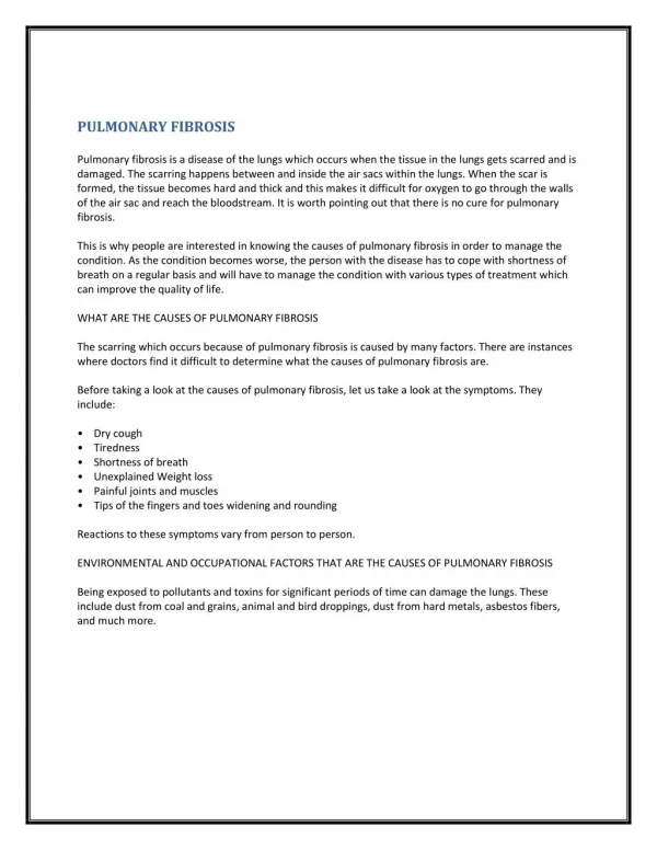
Fibrosis fibrosisis used more broadly to denote is the excessive deposition of collagen and other ECM components in a tissue. As already mentioned, the terms scar and fibrosis are used interchangeably, but fibrosis most often indicates the deposition of collagen in chronic inflammatory diseases (cirrhosis, chronic pancreatitis,pulmonary fibrosis). The basic mechanisms that occur in the development of fibrosis associated with chronic inflammatory diseases are generally similar to the mechanisms of skin wound healing. However, in contrast to the short-lived stimulus that triggers the orderly steps of wound healing in the skin, the injurious stimulus caused by infections, autoimmune reactions, trauma, and other types of tissue injury persists in chronic diseases, causing organ dysfunction and often organ failure.