Download
1 / 70
760 likes | 1.03k Views
Section 3 Fruit tree stem and root disease. 苹果树皮腐烂病 Valsa mali. 苹果黑腐病. 梨树腐烂病 Valsa ambiens. 梨枝干轮纹病 - 粗皮病 Physalospora piricola. 梨干腐病 Botryosphaeria dothidea. 苹果根朽病 Apple root rot. Armillariella tabescens. Fruit Root Cancer 果树根癌病. Dry rot 干腐病. Ring disease. 桃树流胶病 Peach bleeding.

E N D
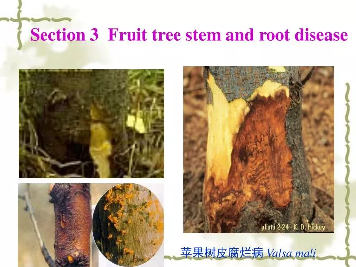
Section 3 Fruit tree stem and root disease 苹果树皮腐烂病 Valsa mali
苹果根朽病 Apple root rot
Fruit Root Cancer 果树根癌病
Dry rot 干腐病 Ring disease
桃树流胶病 Peach bleeding
1 Apple Canker(苹果树皮腐烂病) • 1.1 Occurring and damage • Canker disease mostly occurs on somefruit trees such as apple, peach, and pear, etc. It causes dieback of twigs and branches, • reduces tree growth, causes significant yield losses on fruit trees. Sometimes, it kills the entire tree if the canker girdles the trunk of a tree. • 俗称臭皮病、烂皮病,是我国北方苹果产区危害严重的病害之一。该病主要发生在成龄结果树上,重病果园常常是病疤累累,枝干残缺不全,因病毁园现象时有发生。
1.2 Symptom • Canker by definition means a definite and localized area that is usually dry and dead, often discolored, sunken or raised, sometimes cracked on a stem(主干), trunk(主枝), branch(分枝), or even twig(小枝). • 1.2.1 Onstem:In the early stage of the infection, abnormal appearance may not be visible or identifiable on the bark(树皮) surface. However, when the surface tissue is scraped off, significant discoloration may be seen inside the bark tissue. As a defensive response, tree plants may exude gum from bark at the site of infection or injury.
Bark killed by fungi may be cracked. Some cankered bark may look sooty such as sooty bark canker(溃疡). 1.2.2 On Twigs: the younger branches or twigs may dieback. Sometimes, an entire tree will die if the canker girdles the major stem of a tree. In most cases, fruiting bodies of the fungal pathogen are very evident and visible on dead or seriously infected bark. The most common fruiting bodies are stromata(子座), sporodochia(分生孢子器), and acervuli(子囊壳).
1.3 Pathogen 1.3.1 scientific name and taxon Sexual stage:Valsa mali(有性态为苹果黑腐皮壳),belong to Ascomycotina(囊菌亚门); Asexual stage: Cytospora sp(无性态为壳囊孢),belong to Deuteromycotina(半知菌亚门).
图8-1 苹果树腐烂病菌 左:1.着生于子座组织内的子囊壳 2子囊 3.子囊孢子 右:1.子座剖面示子囊壳 2.子囊壳和子囊孢子 1.3.2. Shape Apple Canker left:1. acervulium子囊壳 2Ascus子囊 3.Ascospore子囊孢子 右:1.子座剖面示子囊壳 2.子囊壳和子囊孢子
1.4 Etiology(病因) Generally speaking, canker disease always occurs on trees that are predisposed by other factors(因素). There are numerous factors that can hurt or stress the tree, forexample, lightening damage, cold and freezing injury, sun scalds, drought, poor soil, nutrient eficiency, improper pruning, other fungal diseases, wild animal damage, and bark borer insects.
1.5 Diseases Cycle Most canker-causing fungi overwinter in dead or infected bark tissue in which fungal fruiting bodies, spores or mycelia (菌丝体)are present. In the spring, fungal spores are transmitted by wind, rain, water, or pruning(修枝) tools to other trees or other parts of the same tree. If spores reach a stressed or injured tree and find desirable infection sites (usually those injured spots or natural holes on the bark), they germinate immediately and then penetrate into the bark tissue. Once the fungus is established in the bark tissue, it kills bark cells and reproduces asexual or sexual fruiting structures(有性或无性繁殖体)there.
During summer time, the fungus usually produces conidia (分生孢子)(asexual state) that can be spread to other trees and start another cycle of infection. This cycle may occur many times during the growing season. When winter is approaching, the fungus will turn into sexual eproduction and produce ascospores.(子囊孢子)Ascospores infect trees in the next spring, resultingin primary infection.
1.6 Control (防治方法 ) • Management of canker diseases should be based on integrated pest management(IPM) principles. • As most of the canker fungi are opportunistic and like to attack those weakened trees, keeping tree’s growth vigor is very critical to lower the chance ofinfection. Avoiding wounding or severe pruning also will reduce opportunities for fungi to attack.
Removing cankers and treating with a disinfectant will prevent further expanding of the canker area. Burning or burying all cankered or dead branches or twigs will eliminate the infection sources. • Always spray some protective fungicides after pruning and before rain.
2.1 Occurring and damage The disease gains its name from the large tumour-like swellings (galls) that typically occur at the crown of the plant, just above soil level. Although it reduces the marketability of nursery stock, it usually does not cause serious damage to older plants. Nevertheless, this disease is one of the most widely known, because of its remarkable biology. Basically, the bacterium transfers part of its DNA to the plant, and this DNA integrates into the plant’s genome, causing the production of tumours and associated changes in plant metabolism.
Crown gall is caused by the bacterium Agrobacterium tumefaciens(根癌土壤杆菌). This bacterium has the widest host range of any plant pathogen. It is capable of causing tumors, or “galls”, on virtually all plant species, except the monocots (grasses). A similar bacterium, Agrobacterium rubi(悬钩子土壤杆菌), causes galls on the canes of brambles(悬钩子). The disease is particularly destructive on brambles (raspberries(悬钩子) and blackberries(黑莓)) and grapes.
It can also cause severe problems on apple, pear, blueberry(越桔), all stone fruits and on ornamentals. The bacteria induce galls or tumors on the roots, crowns, trunks and canes of infected plants. These galls interfere with water and nutrient flow in the plants. Seriously infected plants may become weakened, stunted and unproductive.
Figure A. Large gall formed at the base of the stem of a rose bush. Figure B. A series of galls (arrowheads) along a branch of a grapevine
2.2 Symptom The name describes the rough galls that develop at the crown--the point at the soil line where the main roots join the stem. Often many similar galls will be found on the secondary or lateral roots. Galls may form on the main stem or branches some distance up from the soil line.
The disease first appears as small overgrowths or galls on the roots, crown, trunk or canes. Galls usually develop on the crown or trunk of the plant near the soil line or underground on the roots. Above ground or aerial galls may form on canes of brambles and highly susceptible cultivars of grape. Although they can occur, aerial galls are not common on fruit trees.
In early stages of development the galls appear as tumor-like swellings that are more or less spherical, white or flesh-colored, rough, spongy (soft) and wart-like. They usually form in late spring or early summer and can be formed each season. As galls age they become dark brown to black, hard, rough, and woody. Some disintegrate(碎裂,分解)with time and others may remain for the life of the plant. The tops of infected plants may appear normal. If infection is severe, plants may be stunted, produce dry, poorly-developed fruit, or show various deficiency symptoms due to impaired uptake and transport of nutrients and water.
2.3. Causal Organism Crown gall is caused by the bacterium, Agrobacterium tumefaciens(. A. tumefaciens is a Gram-negative, non-sporing, motile, rod-shaped bacterium. Cane gall of brambles is caused by a closely related bacterium, Agrobacterium rubi. Some scientists consider both species to be widely distributed in soil. The organisms are capable of surviving in soil for at least a year and possibly longer.
The bacteria can enter the plant only through wounds, and much infection in nurseries is through grafting and budding scars. Mechanical injuries of crown and roots by cultivation equipment, animals, and insects are also important entry points.
The crown gall bacterium is soil-borne and persists for long periods of time in the soil in plant debris. It requires a fresh wound in order to infect and initiate gall formation. Wounds that commonly serve as infection sites are those made during pruning, machinery operations, freezing injury, growth cracks, soil insects and any other factor that causes injury to plant tissues.
Bacteria are abundant in the outer portions of primary galls, which is often sloughed off into the soil. In addition to primary galls, secondary galls may also form around other wounds and on other portions of the plant in the absence of the bacterium. The bacteria overwinter inside the plant (systemically) in galls, or in the soil. When they come in contact with wounded tissue of a susceptible host, they enter the plant and induce gall formation, thus completing the disease cycle. The bacteria are most commonly introduced into a planting site on or in planting material.
2.4. The infection process Agrobacterium tumefaciens is found commonly on and around root surfaces - the region termed the rhizosphere - where it seems to survive by using nutrients that leak from the root tissues. But it infects only through wound sites, either naturally occurring or caused by transplanting of seedlings and nursery stock. This requirement for wounds can be demonstrated easily in laboratory conditions.
Figure C shows the bases of two young tomato plants where a drop of A. tumefaciens bacterial suspension was placed on the stem and a pin prick was then made into the stem at this point. The photograph was taken 5 weeks later. Figure D shows another laboratory assay, where bacterial suspension was added to the surface of freshly cut carrot disks。After 2 weeks the young galls (green-coloured) developed from the meristematic tissues(分生组织) around the central vascular system.
In natural conditions, the motile cells(游动细胞) of A. tumefaciens are attracted to wound sites by chemotaxis(趋化性). This is partly a response to the release of sugars and other common root components, and it is found even in plasmid-cured strains. However, strains that contain the Ti plasmid respond even more strongly, because they recognise wound phenolic compounds such as acetosyringone(乙酰丁香酮)(Figure F) which are strongly attractive at even very low concentrations (10-7 Mol). Thus, one of the functions of the Ti plasmid is to code for additional, specific chemotactic receptors that are inserted in the bacterial membrane and enable the bacterium to recognise wound sites.
Acetosyringone plays a further role in the infection process, because at higher concentrations (about 10-5 to 10-4 Mol) than those that cause chemotaxis it activates the virulence genes (Vir genes) on the Ti plasmid (see Figure G). These genes coordinate(调整)the infection process and, in particular. • lead to the production of proteins (permeases,透性酶) that are inserted in the bacterial cell membrane for uptake of compounds (opines) that will be produced by the tumours. • cause the production of an endonuclease - a restriction enzyme - that excises part of the Ti plasmid termed the T-DNA (transferred DNA).
As shown diagrammatically in Figure E, the excised T-DNA is released by the bacterium and enters the plant cells, where it integrates into the plant chromosomes and dictates the functioning of those cells. The actual mechanism of transfer is still unclear, but it seems to require a conditioning process, perhaps mediated by the production of cytokinins(细胞分裂素)(plant hormones) by the bacterium. The tzs (transzeatin) gene(反-玉米素合成酶基因)on the Ti plasmid codes for the hormone (Fig. G).
Figure E. Overview of infection of a plant wound site by Agrobacterium tumefaciens. The Ti plasmid codes for a nutrient-uptake protein (opine permease) that inserts in the bacterial cell membrane. The plasmid also copies and excises part of its DNA, which enters the plant cells and causes them to produce opines. Figure F. Structure of acetosyringone.Figure G. Diagram of some major regions of the Ti plasmid of A. tumefaciens strain C58。
T-DNA = transferred DNA; Noc = nopaline catabolising genes; Ori = origin of replication of the plasmid; Con = region governing conjugative transfer of the plasmid to other Agrobacterium strains; Acc = agrocinopine catabolising genes; tzs = transzeatin synthesis; Vir = virulence genes.
It is important to note that only a small part of the plasmid (the T-DNA) enters the plant; the rest of the plasmid remains in the bacterium to serve futher roles. • When integrated into the plant genome, the genes on the T-DNA code for: • production of cytokinins • production ofindoleacetic acid(吲哚乙酸) • synthesis and release ofnovel plant metabolites - the opine(冠瘿碱)and agrocinopines(农杆糖酯).
The plant hormones upset the normal balance of cell growth, leading to the production of galls and thus to a nutrient-rich environment for the bacteria. The opines are unique amino acid derivatives, different from normal plant products, and the agrocinopines similarly are unique phosphorylated sugar derivatives. All these compounds can be used by the bacterium as the sole carbon and energy source, and because they are absent from normal plants they provide Agrobacterium with a unique food source that other bacteria cannot use.
The bacterium is, basically, a rhizosphere inhabitant because pathogenic strains of Agrobacterium could only respond rapidly to wound sites if there were an established population in the root zone. But the Ti plasmid is a conjugative plasmid - it can be transferred from one cell to another, under the control of the Con region (Figure G). In laboratory conditions, this conjugative transfer is strongly promoted by the presence of nopaline, so it seems that the pathogenic strain creates the conditions (nopaline(胭脂碱)production from infected wound sites) that enable it to transfer its plasmid to other strains in the rhizosphere.
2.5. Control measures Crown gall is best controlled in orchard and ornamental trees by elimination of infected trees from the nursery. Plants having suspicious swellings at graft unions or near the soil line should be discarded.
Nursery soil in which crown gall has occurred may be treated with a suitable fumigant(熏蒸剂), such as chloropicrin(三氯硝基甲)or methyl bromide(溴化甲烷). Growing a nonsusceptible crop, such as grass, for three years will almost eliminate the organism from the soil.
Sterilizing the grafting and budding tools in a disinfectant solution(消毒液)of 20 percent commercial bleach (漂白剂)or a 1/2 percent solution of potassium permanganate(高锰酸钾)will reduce the spread of bacteria in budding and grafting operations.
Biological control is available for a number of fruit and ornamental crops. This method involves inoculating newly grafted, recently lifted transplants or cuttings with a bacterium that is closely related to the one causing crown gall. This prevents the crown gall bacterium from infecting wounds on the plant. Cultures of this competing bacterium are marketed under the trade name Galltrol. Galltrol can be used on non-food and non-bearing crops.